NF-κB pathway inhibition overview
Do you provide NF-κB pathway inhibition?
How NF-κB pathway inhibition is measured
NF-κB sits upstream of many inflammatory genes. When a stimulus reaches the cell, the transcription factor moves into the nucleus and turns on cytokines and enzymes. Inhibition testing asks whether your ingredient lowers that activation step. Because the genes for IL-6, IL-8, TNF-α and COX-2 all respond to this transcription factor, an effect here can explain a broad calming pattern seen in earlier cytokine work.
Reporter and translocation assays
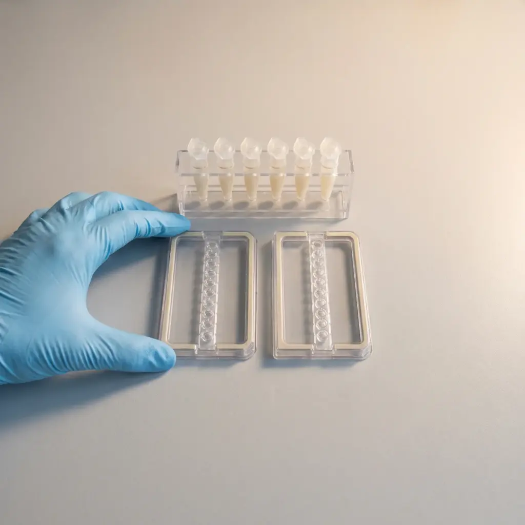
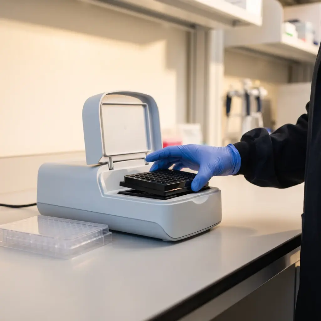
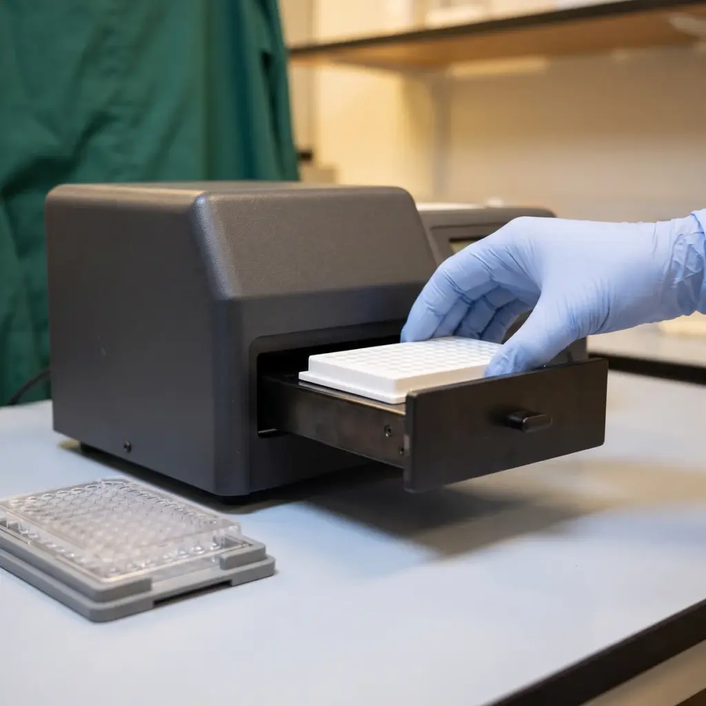
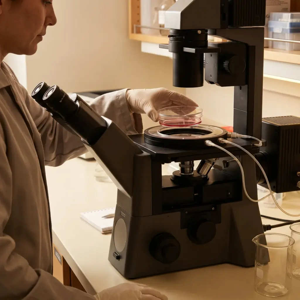
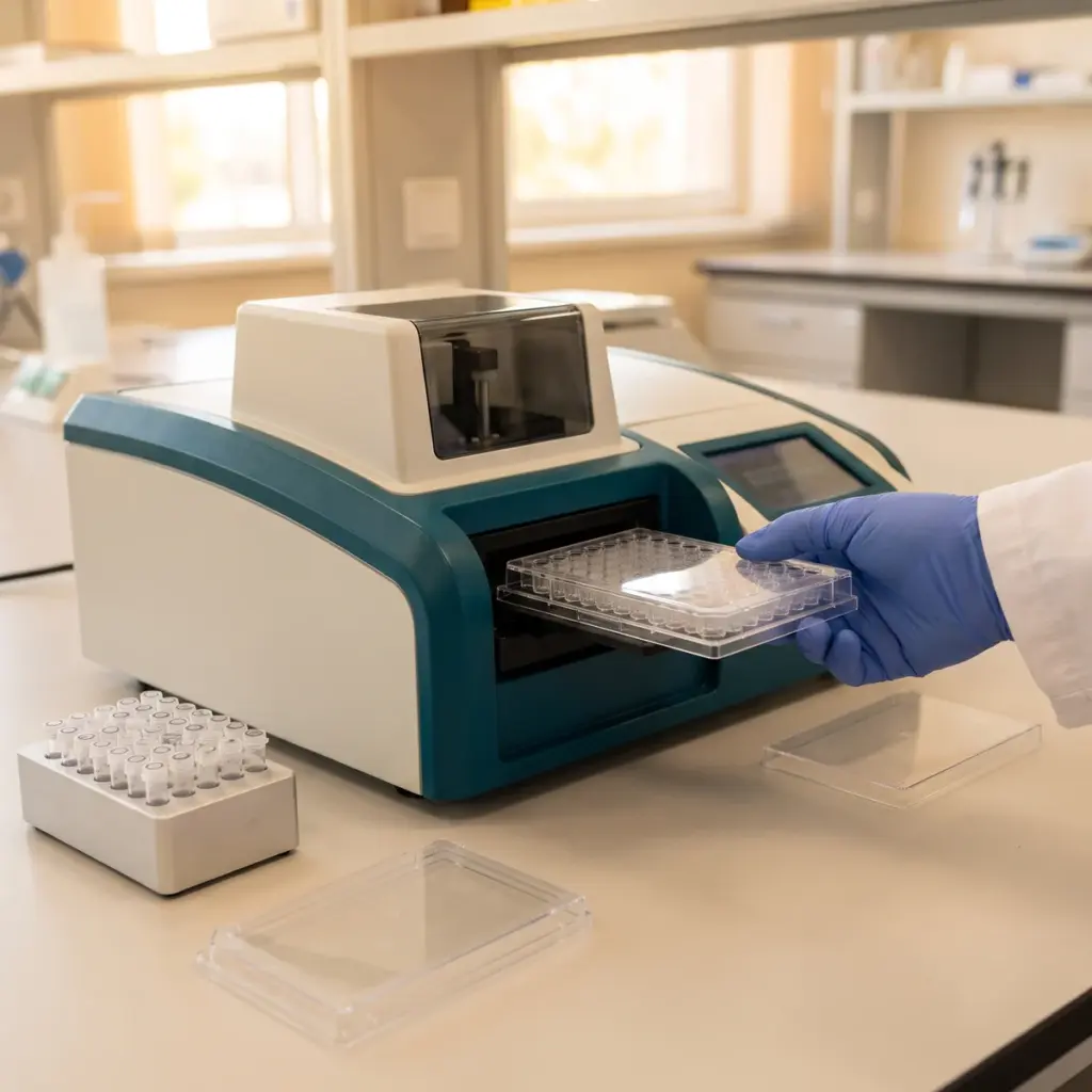
Reporter cell lines carry a construct that produces a light or fluorescent signal when NF-κB is active, which gives a direct, quantitative readout. In keratinocytes and other skin-relevant cells, imaging of nuclear translocation or measurement of pathway proteins adds a second view of the same step. The stimulus, often TNF-α or a bacterial component such as lipopolysaccharide, is chosen to suit the model. Western blotting or immunofluorescence can follow the phosphorylation and breakdown of the inhibitor protein IκBα, the step that releases NF-κB.
- Concentration-response and time-course data
- Stimulated, unstimulated, vehicle and reference controls
- Cell viability results
- Checks for interference with reporter detection
Linking the pathway to output
Downstream measurements of IL-6, IL-1β or COX-2 expression show whether lower signalling carries through to inflammatory output. The TNF-α/IL-6 suppression study covers that output side in more depth. The final report gives you reporter or translocation data, the downstream markers, IC50 values where the curve supports them, and a mechanistic interpretation you can cite in ingredient documentation.
When NF-κB pathway inhibition data earns its place
Commission the study when an active already shows calming behaviour in a cytokine screen and you want to locate where it acts. A cytokine drop alone tells you the output changed. A reporter or translocation result tells you the signalling switch itself moved, which is a stronger basis for a mechanism statement.
What to agree before the quote
Agree the stimulus, the cell model and the readout. A reporter line gives fast, ranked results across many concentrations, while translocation imaging in keratinocytes is closer to skin biology. Ask for viability and interference data with every run, so a quenched signal or a stressed culture is never read as inhibition.
Validated NF-κB reporter lines to look for
Look for a team that has validated its reporter line with a known reference inhibitor and can show reproducible windows between stimulated and unstimulated wells. If you plan volunteer work later, a provider that also runs sensitive skin and skin comfort efficacy testing can carry the mechanism through to a visible redness or comfort endpoint.
More in Inflammation & Skin Immunity
Inflammation & Skin Immunity studies measure how an ingredient changes cytokine, neurogenic and innate defence responses in skin cells and tissue models.