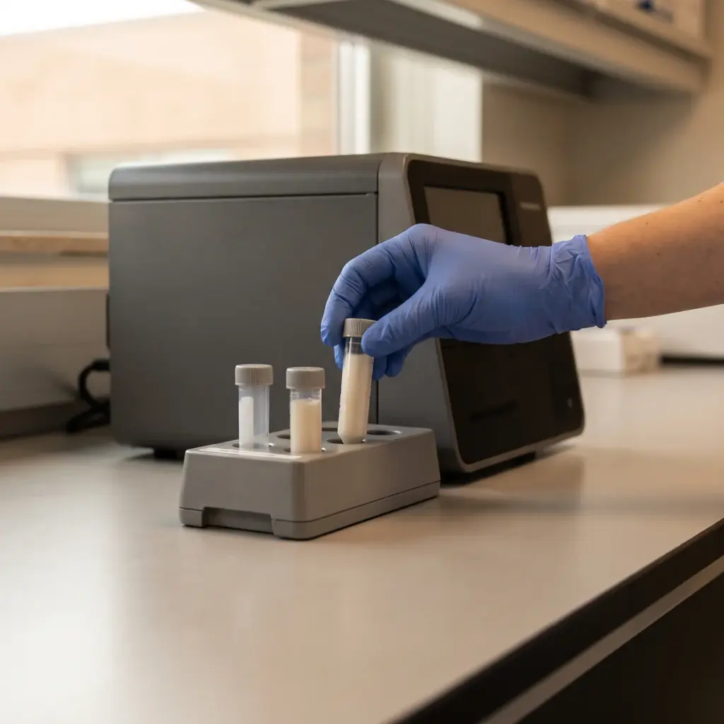
Innate response activation overview
Do you provide Innate response activation?
Measuring innate response activation in skin and immune cells
Innate immunity is the fast layer of skin defence. Cells recognise conserved microbial structures and danger signals released by damaged cells, switch on NF-κB and related transcription factors, and within hours produce cytokines, chemokines and antimicrobial peptides. An activation study maps how strongly and how quickly a test material starts this programme.
Cell systems
Normal human keratinocytes represent the epidermis. Monocytic cell lines such as THP-1, or monocyte-derived macrophages, show how professional immune cells react. Reconstructed epidermis allows topical application of finished formulas. Many studies run a keratinocyte model and an immune-cell model in parallel to compare the two compartments.
Reporter activity, cytokine release and peptide expression
- NF-κB or interferon reporter activity
- IL-1α, IL-1β, IL-6, IL-8 and TNF-α release
- β-defensin and LL-37 gene expression
- Oxidative stress and heat-shock markers
- Cell viability and metabolic activity
Time course and controls
Early transcription changes are read by qPCR within a few hours, and secreted proteins are measured later in the culture medium. Known agonists, such as lipopolysaccharide or a synthetic lipopeptide, are the positive controls and define the maximum response of the system. Your material is then expressed as a fold change against vehicle and as a fraction of that reference activation.
The final report gives the kinetic profile for each marker, dose-response curves and viability data, so you can see which defence pathways the ingredient engages and at what concentration.
Putting innate response activation results to work
An activation study gives a broad first picture of what an ingredient does to skin defence signalling. How you read and extend it depends on the product you are building.
Reading a balanced activation profile
The most useful result for a defence-priming ingredient is a rise in antimicrobial peptides and protective signals with a modest change in inflammatory cytokines. Ask the laboratory to report both groups on the same plate and at the same concentrations, so you can show the ratio. This profile is what separates a defence-supporting active from a simple irritant, and it is the evidence a technical buyer will look for.
Dose finding for formulation
Activation studies give a concentration-response curve. Formulators use it to pick a use level inside the active range and well below concentrations that stress the cells. If the ingredient will be sold in several grades or from several batches, test each one in the same run to document batch consistency.
Following up with targeted studies
Once the activation profile is clear, narrower studies give sharper evidence for a single pathway. TLR modulation testing identifies which receptor the ingredient acts on, and β-defensin expression testing quantifies the peptide response in detail.
More in Inflammation & Skin Immunity
Inflammation & Skin Immunity studies measure how an ingredient changes cytokine, neurogenic and innate defence responses in skin cells and tissue models.