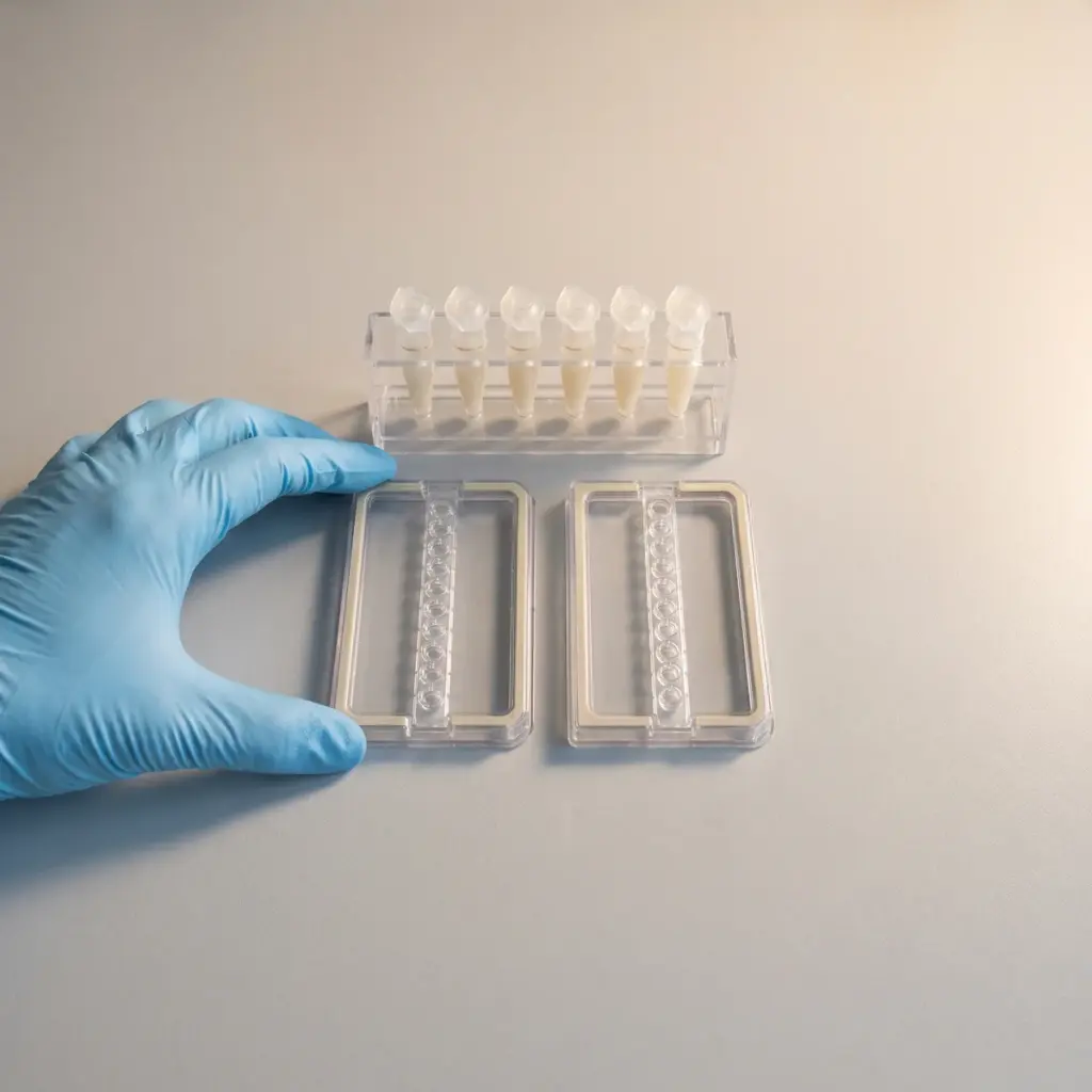
TNF-α/IL-6 suppression overview
Do you provide TNF-α/IL-6 suppression?
Inside a TNF-α/IL-6 suppression study
Tumour necrosis factor-alpha and interleukin-6 are among the first signals a stressed skin or immune cell releases. A suppression study concentrates on these two cytokines, which keeps the design tight and the result easy to compare between candidates. TNF-α acts early and amplifies the response, while IL-6 rises later and sustains it, so the pair covers both the start and the spread of inflammation.
Cell models and stimuli
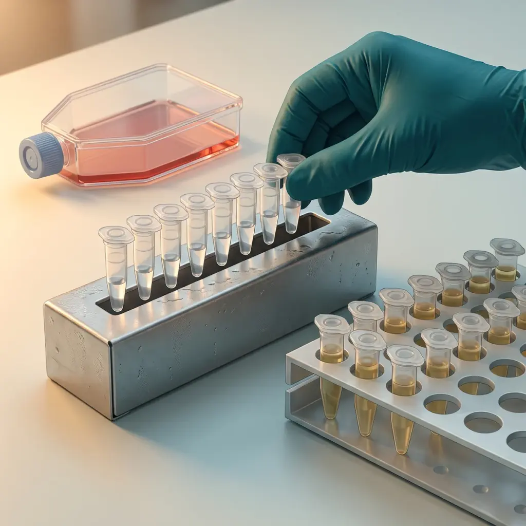
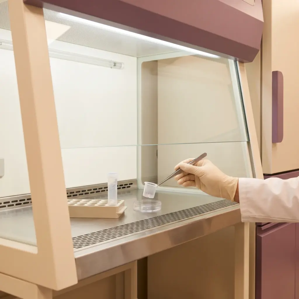
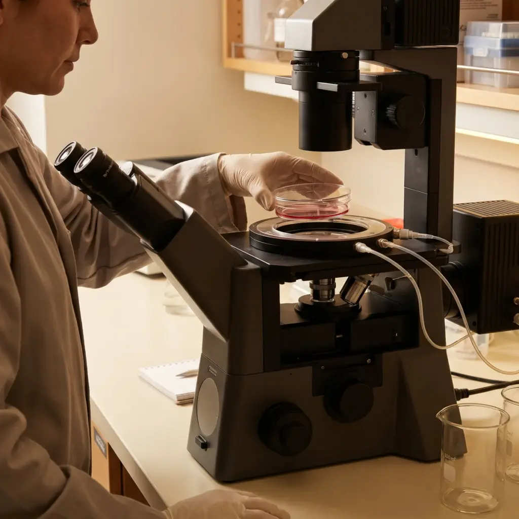
Keratinocytes, macrophages or other immune cells are chosen for a strong, reproducible response to the stimulus. The cells receive the stimulus with and without the test material across a concentration range, and samples are taken at set time points. Common stimuli include lipopolysaccharide for macrophages and a cytokine mix or irritant for keratinocytes.
Assays and controls
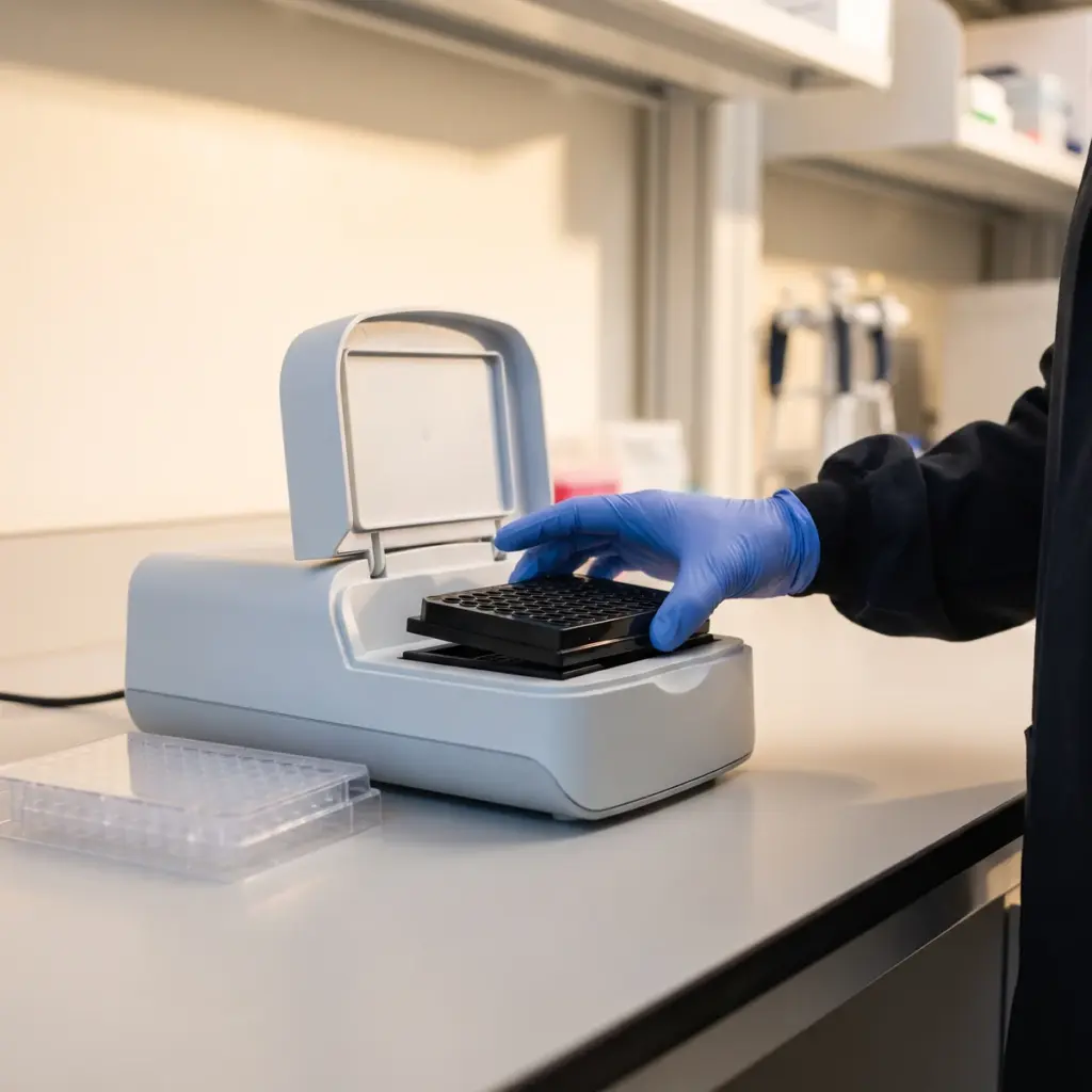
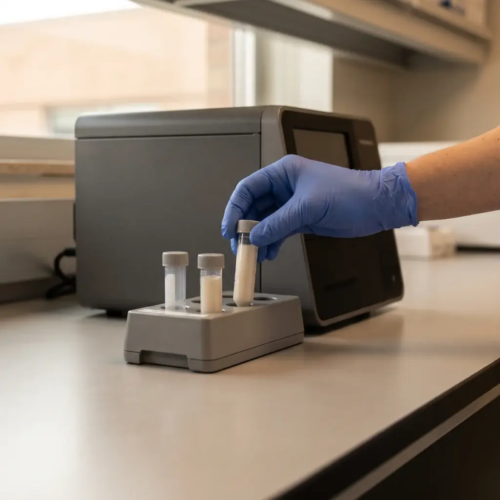
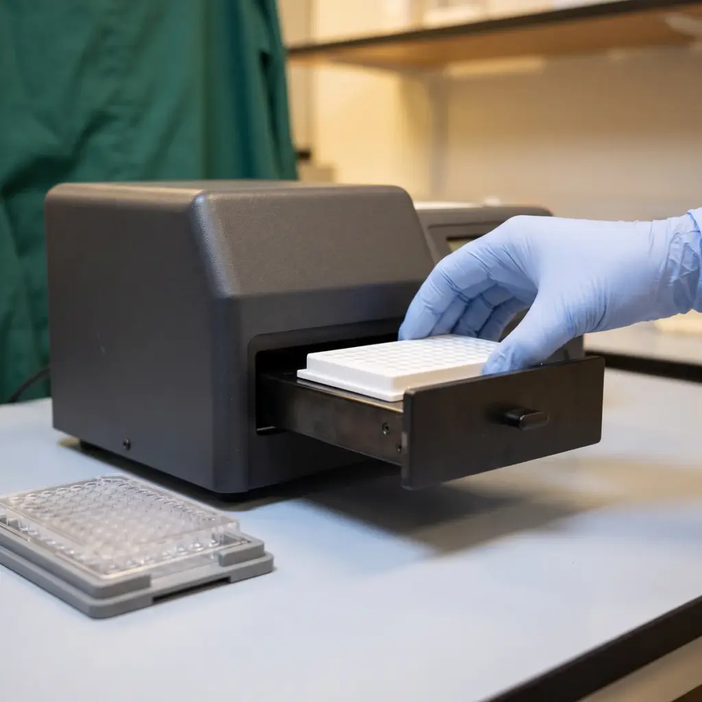
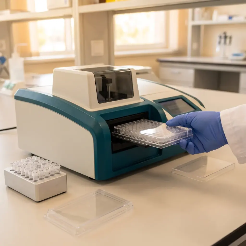
ELISA or multiplex immunoassays measure TNF-α and IL-6 protein in the culture medium. Quantitative PCR measures the matching transcripts, which shows whether the effect starts at gene level. IL-1β can be added when it gives useful context.
- Stimulated and unstimulated wells
- Vehicle and reference-compound comparisons
- Concentration-response and time-course analysis
- Viability and cell-number checks
Cytokine concentrations, IC50 values and viability reported
You receive cytokine concentrations for every condition, percentage change against the stimulated control, IC50 values where the data allow, viability results and a short discussion. Protein and transcript results sit in separate tables, and the discussion addresses cell number and challenge delivery so the suppression figure stands on firm ground.
Using TNF-α/IL-6 suppression results
This is a compact study with a clear numerical output, which makes it one of the easier in vitro results to put into a technical data sheet or a sales presentation for a soothing active. Plan how you will use the figure before you fix the design.
Screening and ranking
Two markers and a fixed stimulus make this a practical screen for several candidate actives or concentrations. The ranking shows which material gives the strongest cytokine drop at a usable concentration, and that becomes the formulation starting point.
Building towards a claim
A clear in vitro reduction supports a mechanism statement in ingredient literature and gives a rationale for a later volunteer study on redness or comfort. Barrier measures such as transepidermal water loss and patch-test tolerance belong to separate studies, so plan them alongside. If you want to show where in the signalling chain the active works, add NF-κB pathway inhibition testing. For a wider view of mediators, a cytokine profile modulation panel reads more signals at once.
More in Inflammation & Skin Immunity
Inflammation & Skin Immunity studies measure how an ingredient changes cytokine, neurogenic and innate defence responses in skin cells and tissue models.