Dendritic cell function overview
Do you provide Dendritic cell function?
Dendritic cell function: activation markers, cytokines and models
Dendritic cells capture material that crosses the skin barrier and carry it to the lymph nodes, where they present it to T cells. When they meet a sensitiser or an inflammatory signal, they mature: they raise co-stimulatory molecules on their surface, change their cytokine output and begin to migrate. Measuring these changes shows how a test material acts on this cell type.
Cell line activation assays
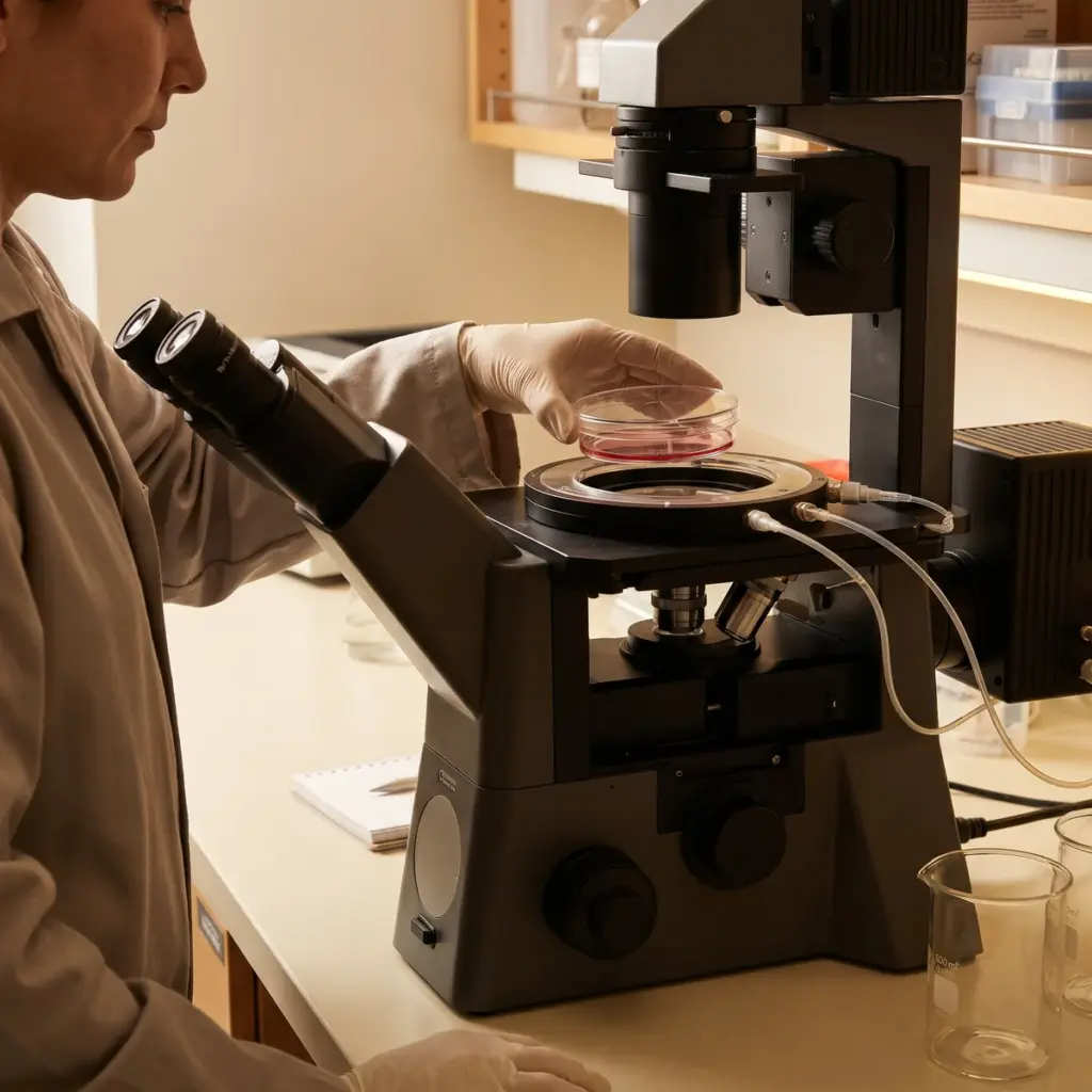
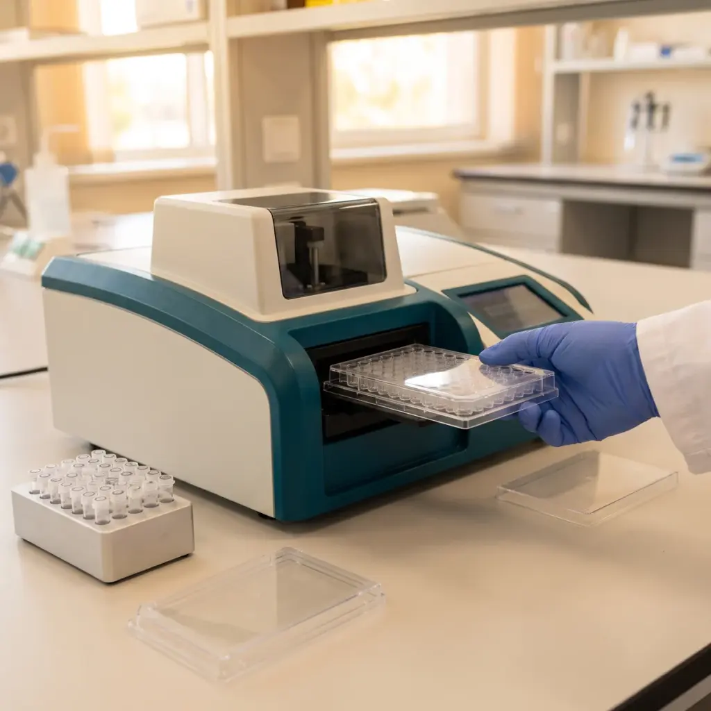
The human Cell Line Activation Test (h-CLAT) exposes THP-1 monocytic cells to a range of concentrations and measures CD86 and CD54 expression by flow cytometry. It is one of the methods in OECD Test Guideline 442E, which covers in-vitro assays addressing dendritic cell activation in skin sensitisation. These methods have defined prediction models and acceptance criteria, and they run to the guideline protocol.
Primary dendritic cell studies
Monocyte-derived dendritic cells, generated from donor blood, give a closer picture of human cell behaviour for exploratory work. Laboratories measure CD80, CD83, CD86 and HLA-DR, release of IL-12, IL-10, TNF-α and IL-8, and the ability to stimulate T cells in co-culture. Langerhans-like cells can also be integrated into reconstructed skin for topical application studies.
Marker fluorescence, cytokines and prediction model classification
- Marker expression as relative fluorescence intensity against vehicle
- Cytokine concentrations per condition
- Cytotoxicity data used to set test concentrations
- A classification against the method’s prediction model, for guideline assays
Using dendritic cell function studies for safety and efficacy
The same cell type answers a safety question and an efficacy question, and the study design follows from which one you are asking.
Sensitisation assessment of new ingredients
A guideline dendritic cell assay addresses one step of the skin sensitisation pathway. It is usually combined with a peptide reactivity assay and a keratinocyte activation assay, and the three results are interpreted together in a defined approach. If you are introducing a new raw material, plan the three studies as a set and ask the laboratory whether it runs all of them under GLP or to the guideline protocols.
Immune-calming efficacy work
For an active presented as calming skin immunity, primary dendritic cells are challenged with a TLR agonist or a known sensitiser, with and without the ingredient. Lower CD86 and IL-12 after treatment show that the active holds the cells in a less activated state. This sits well beside cytokine profile modulation data from keratinocytes.
Preparing the material
Dendritic cell assays are run in culture medium, so solubility decides what can be tested. Supply the ingredient with its solvent and purity, plus any data on poorly soluble fractions. Mixtures and botanical extracts may need a discussion with the laboratory about the applicability domain of the chosen method before the study starts.
More in Inflammation & Skin Immunity
Inflammation & Skin Immunity studies measure how an ingredient changes cytokine, neurogenic and innate defence responses in skin cells and tissue models.