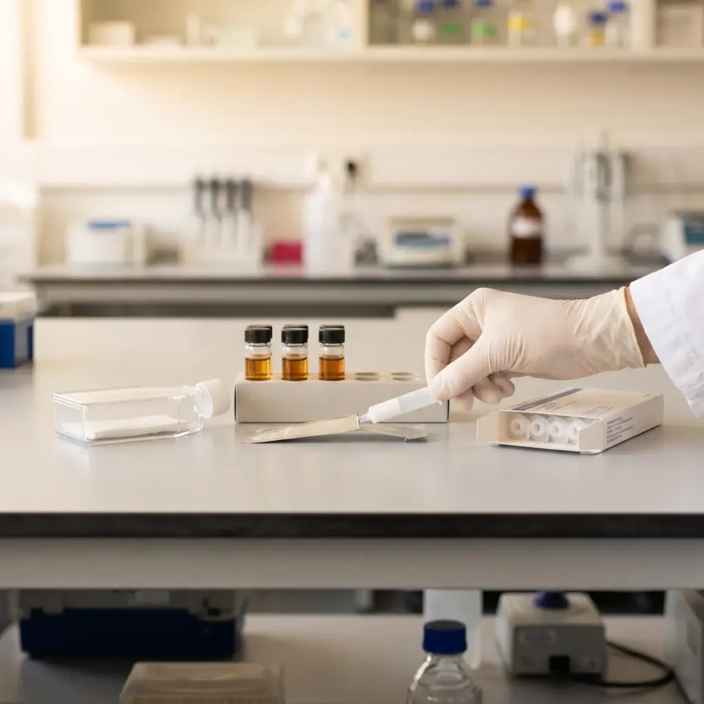
AMP production overview
Do you provide AMP production?
AMP production testing: peptides, models and functional readouts
Antimicrobial peptides are short, mostly cationic molecules that keratinocytes secrete onto the skin surface and into the upper epidermis. They disrupt microbial membranes, shape which bacteria thrive on the skin and send signals to immune cells. Their production rises after injury, microbial contact and certain vitamin and fatty acid signals, which is what an AMP study looks for.
The peptides measured
- Cathelicidin LL-37, encoded by the CAMP gene
- Human β-defensins hBD-1, hBD-2 and hBD-3
- Psoriasin (S100A7)
- RNase 7
- Dermcidin, from sweat gland models where relevant
Keratinocyte and reconstructed epidermis models for AMPs
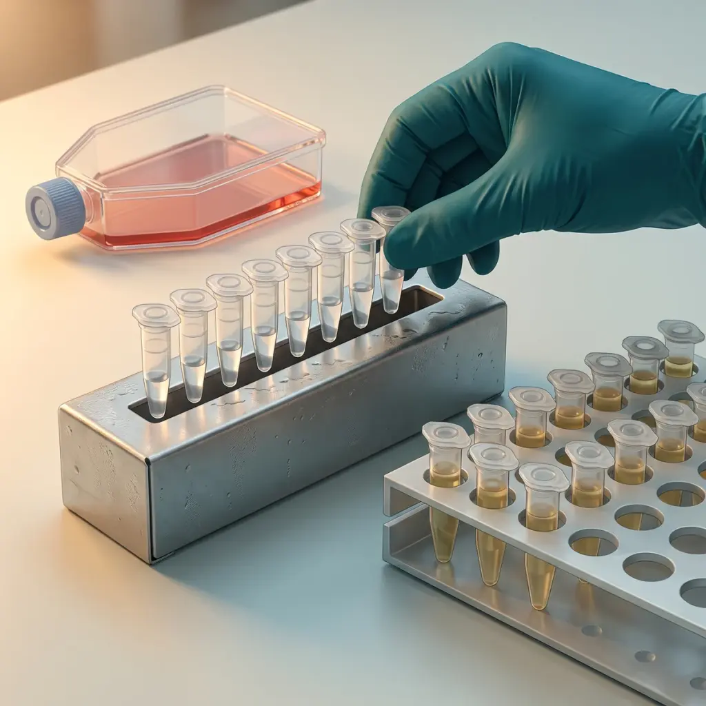
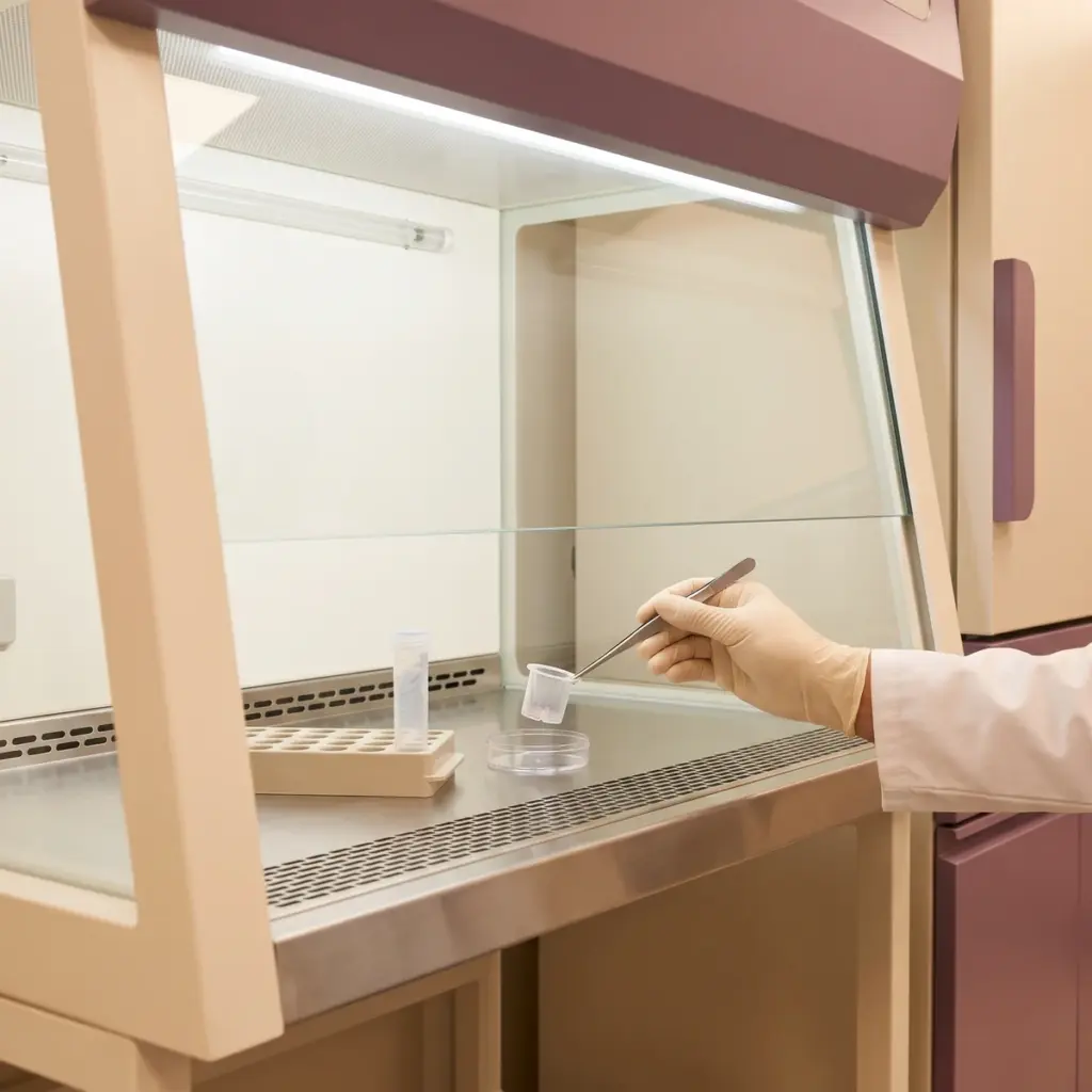
Normal human keratinocytes in monolayer give a fast screen for gene expression. Reconstructed epidermis, which forms a stratum corneum, accepts topical application of creams and serums and secretes peptides into the medium and the tissue. Some protocols add a bacterial stimulus, such as heat-killed skin bacteria, to model the response to microbial contact.
From transcript to protein to activity
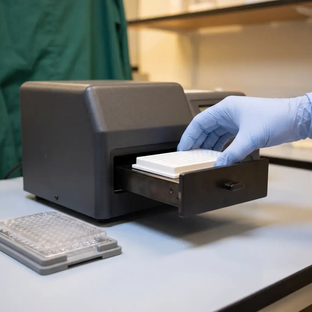
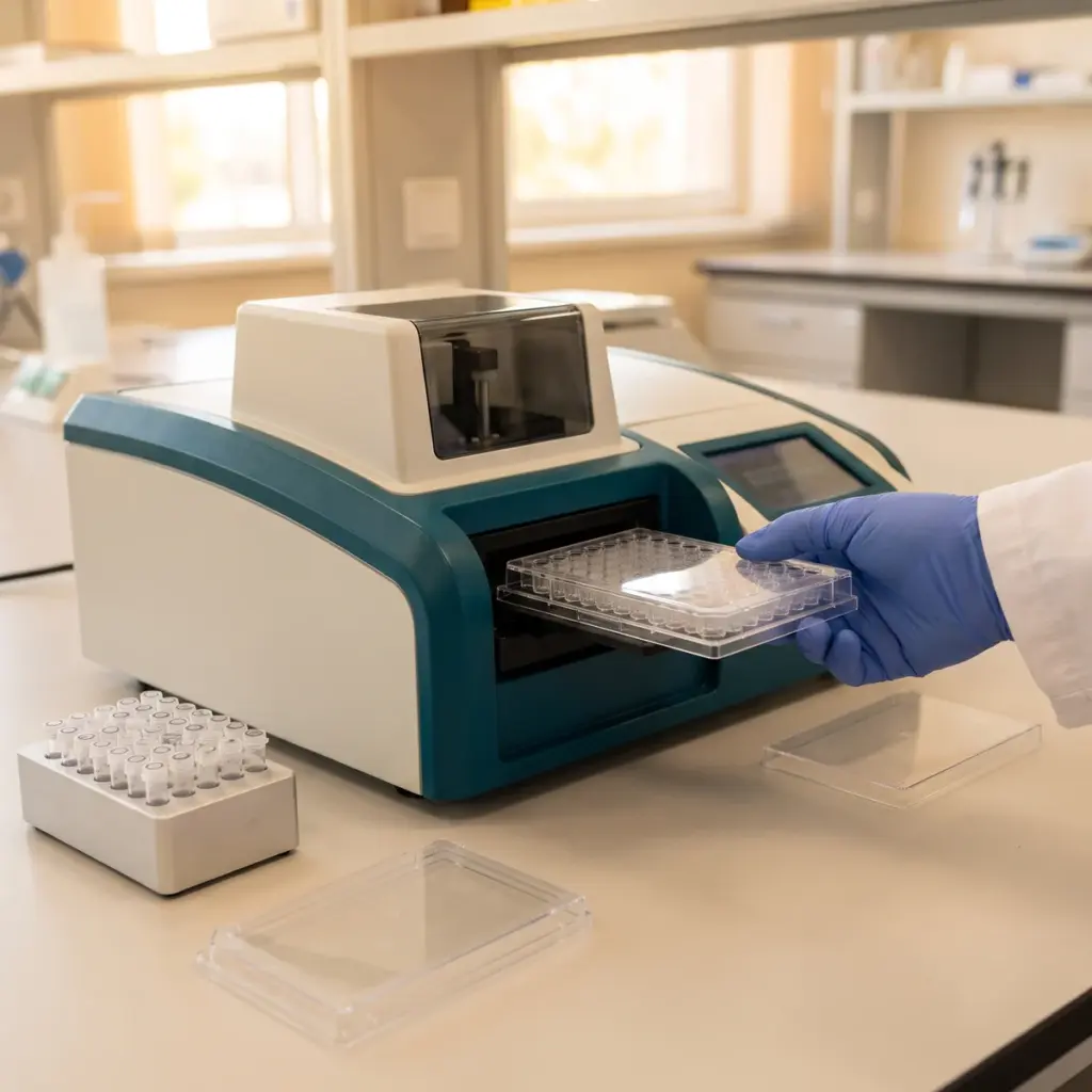
qPCR measures mRNA for each peptide relative to housekeeping genes. ELISA quantifies secreted LL-37, hBD-2 or psoriasin. Immunostaining on tissue sections shows where in the epidermis the peptide accumulates. A functional step, in which conditioned medium is incubated with a reference bacterial strain and growth is measured, connects the peptide increase to antimicrobial activity.
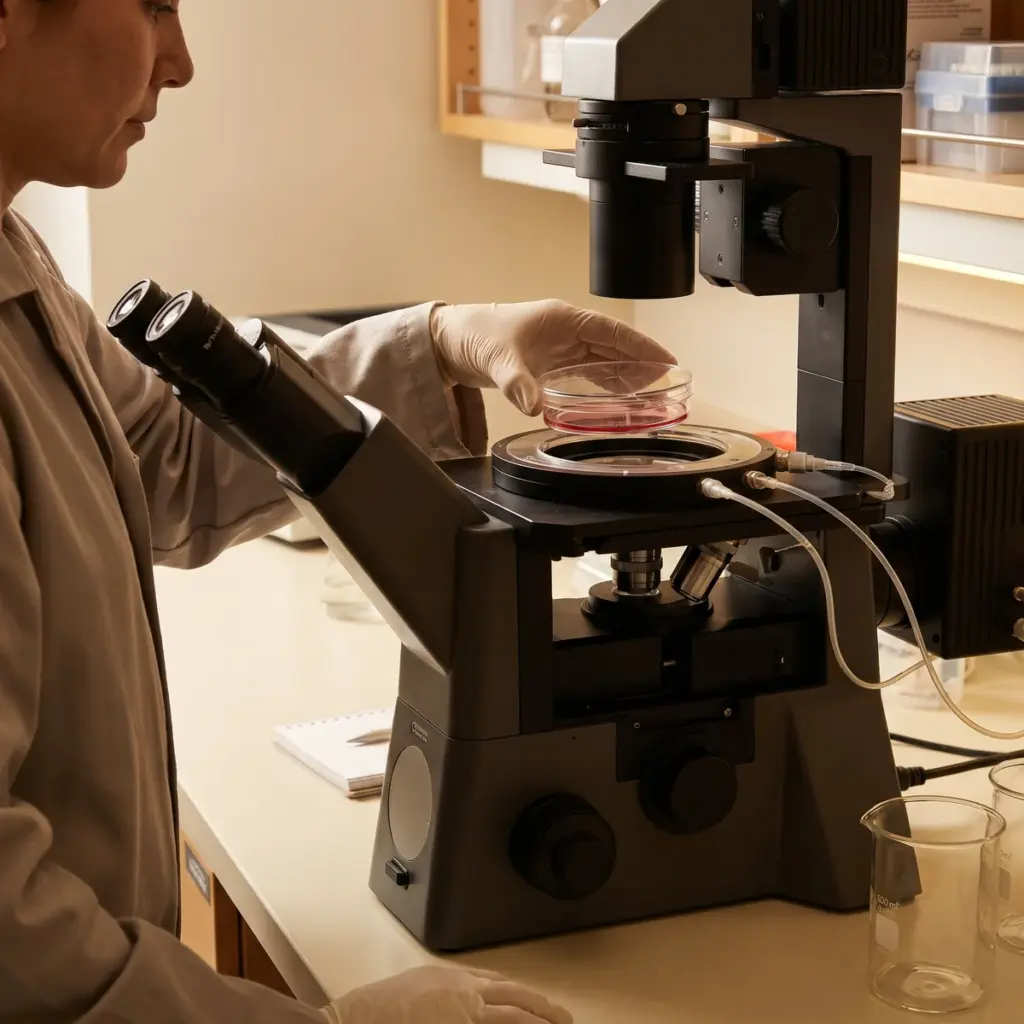
Results are reported as fold change against untreated tissue and a positive control, with viability data and photomicrographs where staining is included.
Applications of AMP production studies for skin defence products
AMP studies are commissioned mostly at the ingredient stage, before a defence or microbiome positioning is fixed for a finished product.
Choosing the peptide panel
Match the panel to the ingredient’s proposed action. Vitamin D-related actives and some fatty acids act mainly on cathelicidin. Bacterial ferments and lysates often raise hBD-2 and psoriasin through TLR signalling. If you are unsure, a broad qPCR screen on keratinocytes shows which peptides respond, and a narrower ELISA study on reconstructed tissue then measures the protein. For a single family studied in depth, see β-defensin expression testing.
Microbiome and defence positioning
AMP data gives a defence or microbiome-balance claim a measurable cellular basis. It pairs well with a volunteer study in which skin swabs are analysed by sequencing, so you can show the cellular mechanism and the change on the skin surface together. Keep the AMP study and any preservative efficacy testing separate in your planning: they answer different questions about different materials.
Product type and antimicrobial step for quoting
Send the ingredient’s form, solvent, use level and origin. For finished formulas, tell the laboratory the product type, because rinse-off and leave-on products need different exposure times on reconstructed tissue. Say whether you need the functional antimicrobial step, since it requires a microbiology laboratory alongside the cell culture team.
More in Inflammation & Skin Immunity
Inflammation & Skin Immunity studies measure how an ingredient changes cytokine, neurogenic and innate defence responses in skin cells and tissue models.