Clinical imaging documentation overview
Do you provide Clinical imaging documentation?
Choosing a clinical imaging method for skin claims
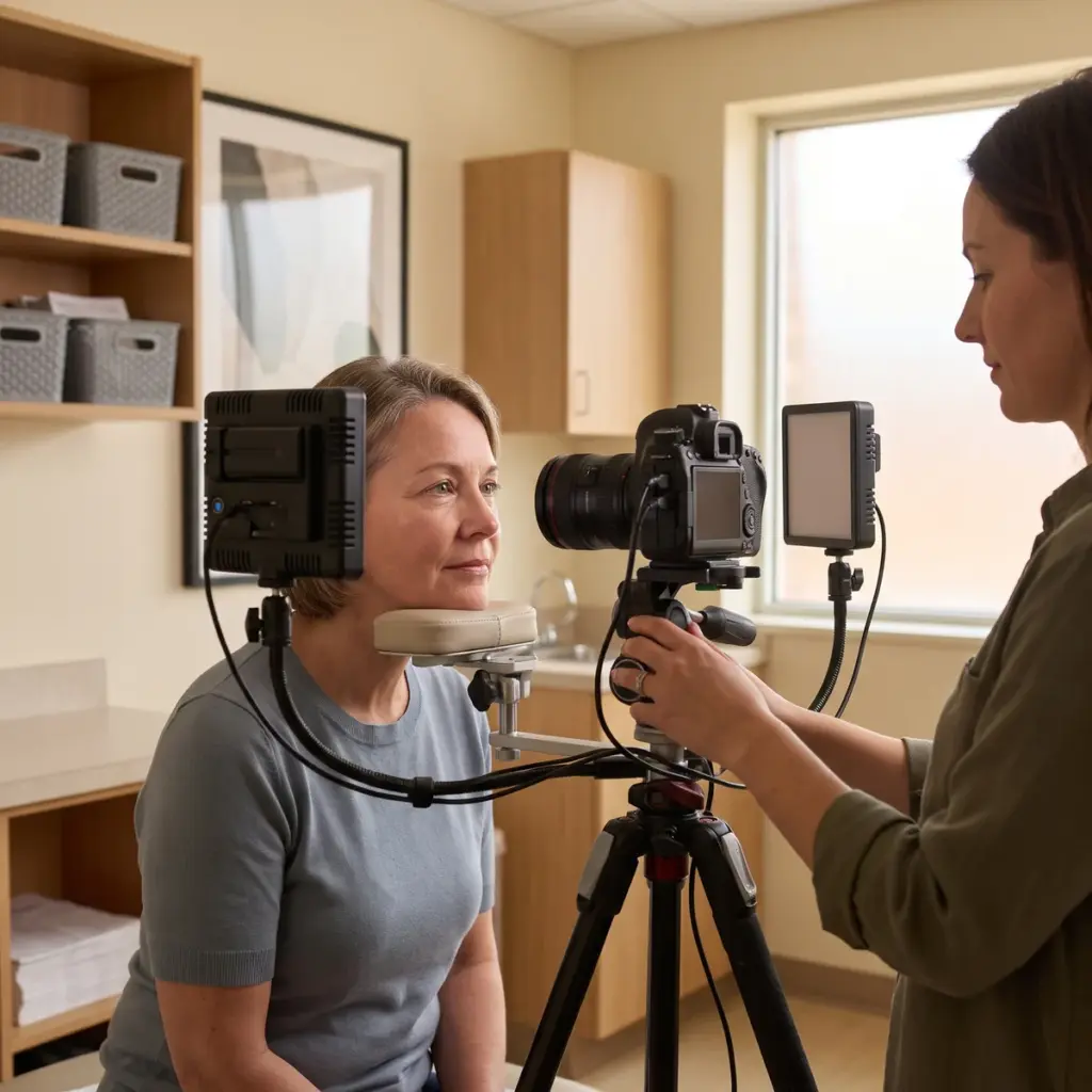
All clinical imaging shares one discipline: every visit must be captured the same way. Lighting, camera settings, distance, head position, facial expression and skin preparation are fixed in the protocol, and alignment aids help reproduce the pose. That consistency is what lets a change between baseline and follow-up be read as a change in the skin. The four methods in this family each measure something different.
Photography in visible and polarised light
Standardised photography produces calibrated colour images of the face or body. Parallel- and cross-polarised modes separate surface shine from colour beneath the surface. Trained graders score the images against reference scales, and image analysis software can count or measure features such as spots or pores.
Ultraviolet imaging
UV photography uses reflected ultraviolet light or UV-induced fluorescence. It shows pigment irregularities that are faint in normal light and, in fluorescence mode, features such as porphyrins in pores.
Surface relief measurement
3D surface topography scans the skin with projected light patterns and calculates height maps. From these the laboratory derives wrinkle depth, volume and roughness values for statistical comparison between visits.
Magnified and in-depth imaging
Clinical imaging microscopy covers dermoscopy and reflectance confocal microscopy. Dermoscopy magnifies surface detail and follicle openings. Confocal microscopy shows cell-level structure and pigment distribution in the upper skin layers without taking a sample.
Services in Clinical imaging documentation
Planning clinical imaging documentation within an efficacy study
Bring imaging into the study design early. The method follows from the claim: a visible-wrinkle claim points to photography with grading or to 3D topography, a dark-spot claim to cross-polarised or UV photography, and a pore or texture claim to topography or dermoscopy. Many studies combine a photographic record for marketing with an instrumental method for the statistics.
Details to settle before asking for quotes
List the claims, the body sites to image, the number of visits and the endpoints the images must deliver (expert grades, counts, depth values). Say whether you will want images for advertising, since this affects consent wording and the file formats supplied. Ask for the untouched original files as well as any marketing selections.
Evaluating an imaging laboratory
Check which systems the laboratory runs and how often they are calibrated. Ask how they reposition volunteers between visits, how they control ambient light and how they store original files with their metadata. Graders should be trained on a validated scale and blinded to the visit where possible. Ask too how images and derived measurements are handed over, so they slot into the final study report with their capture settings attached.
More in Study Evidence Documentation
Study evidence documentation turns cosmetic test results, images and source records into reports and claim dossiers a reviewer can follow and check.