Tight-junction proteins overview
Do you provide Tight-junction proteins?
How tight-junction protein testing is carried out
Tight junctions form a belt around each cell in the stratum granulosum. Claudins and occludin span the membrane and close the gap between neighbouring cells. ZO-1 sits on the inner side and anchors them to the cytoskeleton. Together they control the passage of water, ions and small molecules between cells, forming a second barrier beneath the stratum corneum.
Keratinocyte inserts and reconstructed epidermis for junction work
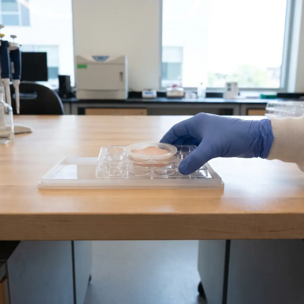
Keratinocyte monolayers grown to confluence on permeable inserts form tight junctions and give a clean system for resistance measurements and dose ranges. Reconstructed human epidermis places the junctions in their natural granular layer position and accepts topical products. Studies can include a challenge, such as an irritant or inflammatory stimulus, to test whether the active keeps junctions intact.
Protein, imaging and functional readouts
- Claudin-1, claudin-4 and occludin levels by Western blot
- ZO-1 and claudin distribution at cell borders by immunofluorescence
- Corresponding transcripts by qPCR
- Transepithelial electrical resistance over time
- Passage of fluorescent tracers of defined size
Imaging matters here because junction proteins must sit at the cell membrane to work. Continuous staining around the cell outline indicates organised junctions, and image analysis can score that continuity. TEER and tracer flux then show whether the seal has tightened. E-cadherin from adherens junctions can be added for a wider picture of cell-to-cell adhesion.
Claudin quantification, confocal images and TEER curves
The report presents protein quantification, confocal images, TEER curves and tracer data for each treatment against vehicle and reference controls, with cell viability and statistics.
Applying tight-junction protein data to barrier claims
Tight junctions give an active a second barrier mechanism, located in the living cell layers. Higher claudin-1 at the cell borders combined with a rise in electrical resistance supports wording such as “reinforces cell-to-cell cohesion in the epidermis” or “strengthens the skin’s inner barrier in a reconstructed model”. The functional data are what make that claim convincing.
Combining junction work with other barrier studies
Many barrier actives act on several levels. A full programme often pairs tight-junction analysis with intercellular lipid order for the stratum corneum and keratinocyte differentiation markers for maturation. Running them on one tissue batch keeps the results comparable.
Choosing a provider for junction studies
Ask whether the lab measures TEER on its own models and what baseline values it typically sees, since a stable baseline is needed to detect a treatment effect. Confirm that immunofluorescence is quantified by image analysis of membrane staining. If your product targets sensitive skin, ask about an irritant or inflammatory challenge arm to test junction protection.
More in Barrier & Hydration Mechanisms
Barrier & Hydration Mechanisms studies show how an ingredient acts on epidermal lipids, barrier proteins and water-handling pathways in skin models.