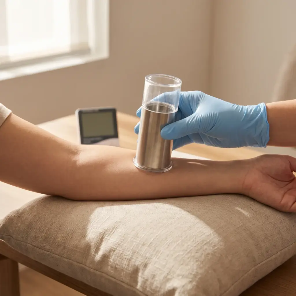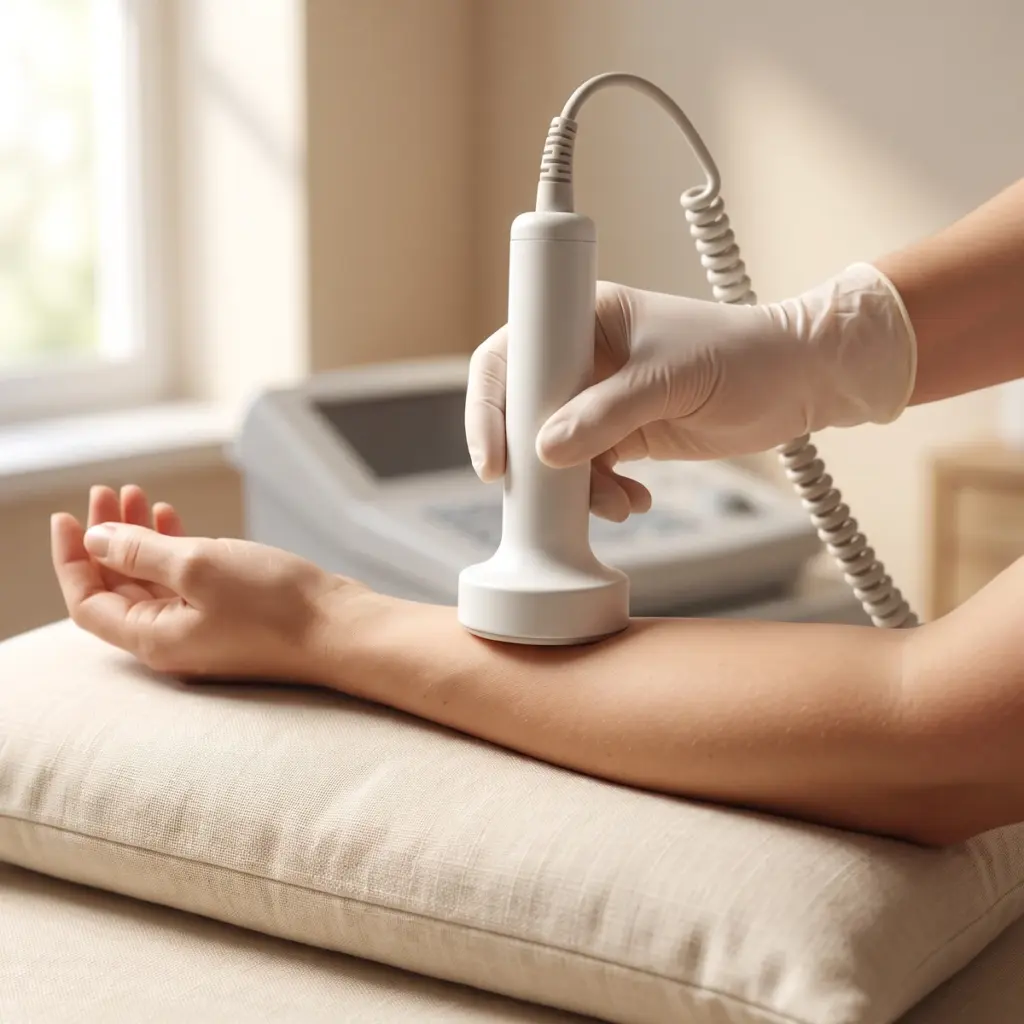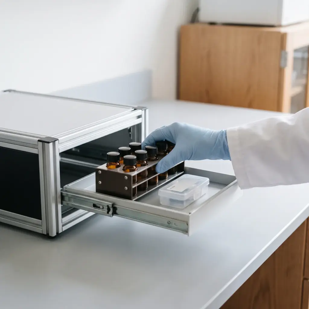
Barrier & Hydration Mechanisms overview
Do you provide Barrier & Hydration Mechanisms?
Barrier & hydration mechanism studies and what each one measures
Most of the skin barrier sits in the stratum corneum, where flattened corneocytes are held in a matrix of ceramides, cholesterol and free fatty acids. Below it, living keratinocytes build that structure as they differentiate, and tight junctions in the granular layer seal the gaps between cells. Water is held by natural moisturising factor (NMF) inside corneocytes and by hyaluronic acid in the living layers, while aquaporin channels move water and glycerol between cells. Each service in this family targets one part of that system.
Epidermal differentiation and barrier proteins
Keratinocyte differentiation studies follow markers such as involucrin, loricrin and transglutaminase. Filaggrin production, calcium gradient maintenance and tight-junction proteins (claudin-1, occludin, ZO-1) are offered as separate studies, read out by gene expression, protein assays or immunostaining of tissue sections.
Stratum corneum lipids
Ceramide synthesis mechanisms and ceramide synthesis in epidermal models quantify ceramide classes, usually by LC-MS. Intercellular lipid order and lamellar lipid reinforcement look at how those lipids pack, with FTIR spectroscopy or X-ray diffraction.
Cohesion and desquamation
Corneodesmosome regulation, proteases inhibition and epidermal cohesion studies measure corneodesmosin, desmoglein-1, kallikrein-related peptidases and their inhibitor LEKTI. Together they describe how an ingredient affects the orderly shedding of corneocytes behind smooth, even skin texture.
Water binding and transport
Hyaluronic acid synthesis, aquaporin expression (mainly AQP3) and NMF production studies measure the molecules that hold and move water. NMF work quantifies amino acids, pyrrolidone carboxylic acid (PCA) and urocanic acid. Each study ends with a report giving the model, concentrations, controls, normalised data and statistics.
Services in Barrier & Hydration Mechanisms
Choosing barrier & hydration mechanism studies for your ingredient
With sixteen mechanism studies on offer, the choice comes down to the claim you want to explain and the model that can show it.
Start from the benefit you want to explain
A humectant-type moisturising active is best characterised with NMF, hyaluronic acid or aquaporin endpoints. An ingredient positioned for barrier strength calls for ceramide, lipid order or tight-junction work. Actives for rough, flaky skin fit cohesion and protease studies. Two or three linked endpoints from one pathway give clearer mechanism evidence than a scattered panel of unrelated markers.
Picking the model
Monolayer keratinocytes are quick to run and suit gene-expression screening of many candidates. Reconstructed epidermis forms a stratum corneum, so it is the model of choice for ceramide profiles, lipid packing and topical application of a finished formula. Ex vivo human skin keeps the native architecture and is used when you need tissue context close to real skin.
Linking mechanism to clinical results
Mechanism data explain why a product works. The measured benefit on volunteers comes from skin hydration efficacy testing with corneometry and from human barrier studies based on transepidermal water loss. A common sequence is to run the mechanism study first to select the lead active, then confirm the finished product on a volunteer panel and present both sets of data in the technical file.
More in Hydration Barrier Testing
Skin hydration and barrier efficacy testing measures moisturisation and water loss on volunteers, with lab studies of the lipids behind the effect.