Filaggrin production overview
Do you provide Filaggrin production?
Filaggrin production assays from gene to NMF
Keratinocytes in the granular layer make profilaggrin, a large precursor stored in keratohyalin granules. During cornification it is cut into filaggrin units that bind and compact keratin filaments. In the upper stratum corneum, filaggrin is then degraded into free amino acids, which are converted in part to pyrrolidone carboxylic acid and urocanic acid. These products make up a large share of natural moisturising factor. A filaggrin study can measure each of these stages.
Epidermal models used
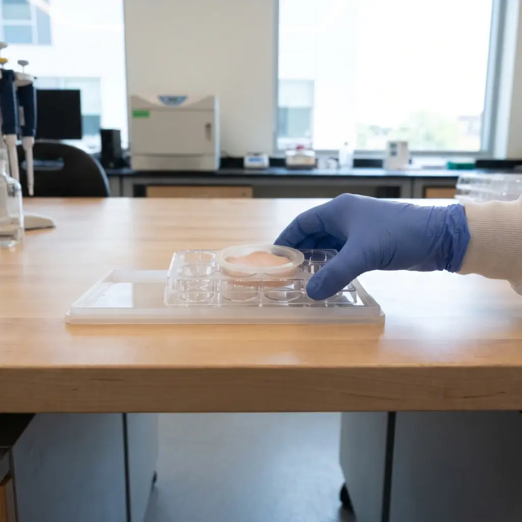
Differentiated keratinocyte monolayers give fast, dose-ranging data on filaggrin expression. Reconstructed human epidermis adds a stratum corneum, which is needed to follow protein processing and NMF release, and it accepts topical application of creams and serums.
Gene, protein and breakdown product readouts
- Filaggrin mRNA by qPCR
- Profilaggrin and filaggrin protein by Western blot
- Localisation in the granular layer by immunostaining
- Free amino acids, PCA and urocanic acid by HPLC or LC-MS
- Tissue viability
Measuring all three levels shows whether an active increases synthesis, changes processing, or both. A rise in transcript with more stained granules and higher NMF in the stratum corneum is the strongest pattern.
Filaggrin immunostaining and NMF results reported
You receive quantified results for each endpoint against untreated and vehicle controls, with statistics, immunostaining images and the full model and dosing description. Where NMF components were analysed, the report lists each measured compound per tissue. The NMF data can be extended in NMF production studies.
Building a hydration claim on filaggrin production data
Filaggrin sits at the junction of barrier structure and water binding, which makes it a strong mechanism for a moisturising active. Results showing more filaggrin and more of its breakdown products support wording such as “boosts filaggrin, a source of the skin’s natural moisturising factor, in reconstructed epidermis”.
Where filaggrin testing fits in a hydration programme
Run it once an active has shown a hydration signal and you need to explain it. The finished product then goes to hydration efficacy testing on volunteers, and the filaggrin data explain the mechanism behind the corneometer readings. Suppliers often combine filaggrin with aquaporin-3 or hyaluronic acid endpoints to describe several water pathways at once.
Details to agree before the study starts
Decide whether you need transcript data only, which is quicker for screening, or the full set with protein and NMF analysis for a claim file. Ask the laboratory which antibody it uses and whether it distinguishes profilaggrin from processed filaggrin on the blot. For NMF analysis, confirm how the stratum corneum is sampled from the tissue and which compounds are quantified. If your formula contains amino acids, PCA or urea, say so, since the lab will need vehicle controls to separate product residues from tissue output.
More in Barrier & Hydration Mechanisms
Barrier & Hydration Mechanisms studies show how an ingredient acts on epidermal lipids, barrier proteins and water-handling pathways in skin models.