Calcium gradient maintenance overview
Do you provide Calcium gradient maintenance?
Mapping the epidermal calcium gradient
In intact epidermis, calcium is low in the basal and spinous layers and highest in the granular layer. This gradient drives the final steps of differentiation and the secretion of lamellar bodies that supply barrier lipids. When the barrier is disrupted, the gradient flattens. It reforms as the barrier recovers, and that recovery is what this service follows.
Tissue models and study design
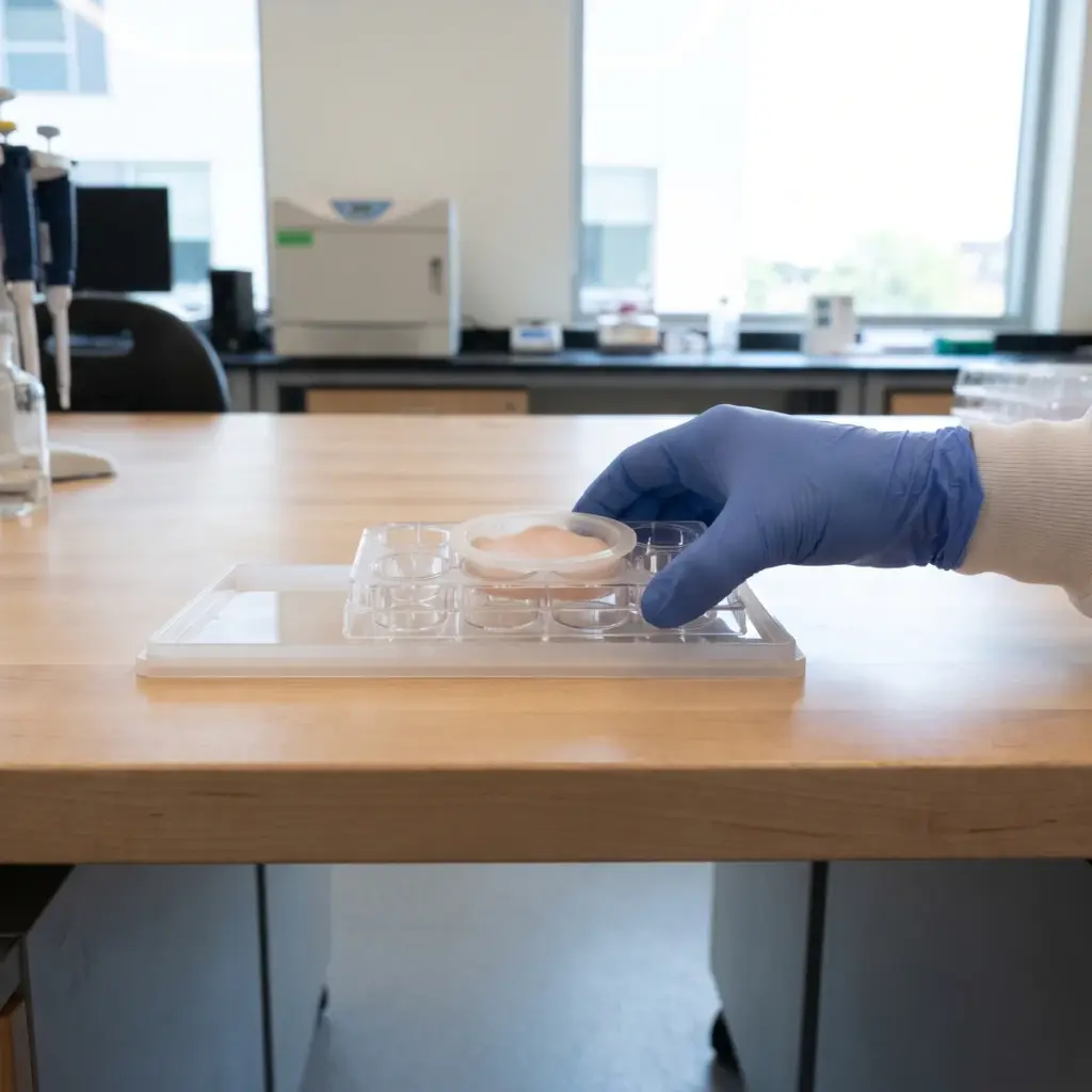
A gradient only exists across a stratified tissue, so studies use reconstructed human epidermis or skin explants. Tissues are treated with the test material, then the barrier is disrupted with a defined challenge such as a surfactant or solvent. Samples are taken at set times during recovery and compared with untreated and vehicle tissues.
Layer-resolved calcium measurement
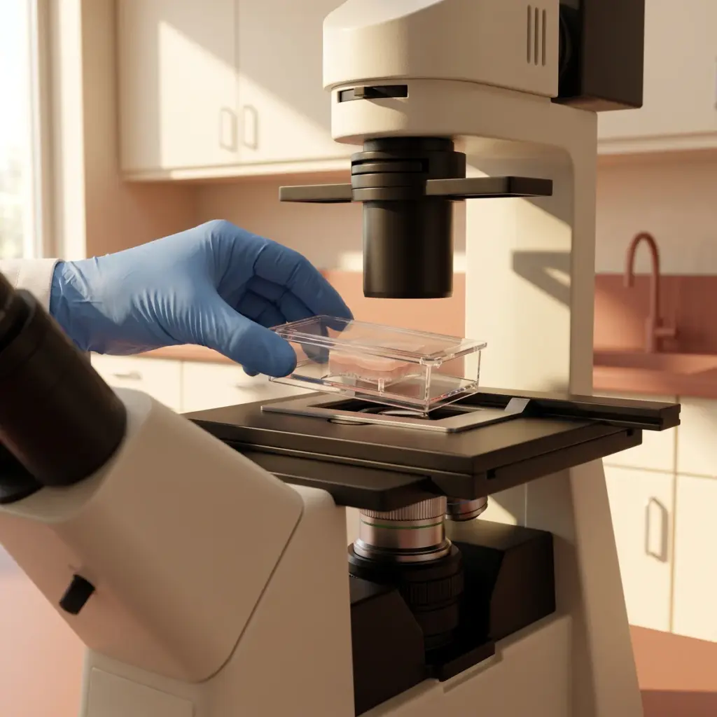
Calcium is localised in tissue sections by histochemical precipitation methods examined under the microscope, or by elemental mapping across the section. The laboratory quantifies the signal layer by layer and plots the profile from basal to granular layer for each group.
- Baseline calcium profile in the model
- Profile loss after barrier disruption
- Recovery of the granular layer peak over time
- Effect of treatment at each time point
Intracellular calcium signalling
Keratinocytes loaded with fluorescent indicators such as Fluo-4 or Fura-2 show how quickly cells respond to an active or to a change in external calcium. These live-cell recordings describe signalling within cells and complement the tissue profile.
The report contains the layer-by-layer profiles, representative images, the signalling traces where included and statistical comparison between groups. Differentiation markers measured on the same tissues, as in keratinocyte differentiation testing, link the calcium result to cell maturation.
Using calcium gradient maintenance data
Calcium mapping answers a specific question about barrier recovery, so it earns its place in a programme once you know your active works and want to show how.
Supporting a barrier recovery claim
A calcium profile that returns to its granular layer peak sooner in treated tissue gives you an upstream mechanism for a barrier recovery claim. It explains why lipid secretion and differentiation restart, which makes the data valuable in an ingredient technical file. Pair it with a functional readout from skin barrier function efficacy testing so the mechanism and the barrier outcome sit in the same dossier.
Fitting it into a test programme
Calcium work usually comes after a first barrier screen has shown that an active speeds recovery. At that point the question becomes how it works, and the gradient is one of the earliest events in barrier repair. It also suits actives that are thought to act on ion channels or calcium-sensing pathways in keratinocytes.
Layer-resolved calcium mapping experience to check
Few laboratories run layer-resolved calcium mapping routinely. Ask to see an example profile from untreated and disrupted tissue in their model, since a clear baseline gradient is the starting point for any treatment effect. Check which disruption method they use and how many recovery time points the design includes, and whether live-cell calcium imaging is available in-house if you want the signalling data too.
More in Barrier & Hydration Mechanisms
Barrier & Hydration Mechanisms studies show how an ingredient acts on epidermal lipids, barrier proteins and water-handling pathways in skin models.