Keratinocyte differentiation overview
Do you provide Keratinocyte differentiation?
Measuring keratinocyte differentiation in culture and tissue models
As keratinocytes leave the basal layer, they switch on a sequence of proteins. Keratins 1 and 10 appear in the spinous layer. Involucrin, transglutaminase-1, loricrin and filaggrin follow in the granular layer, where cells build the cornified envelope and then flatten into corneocytes. A differentiation study measures where along this sequence an active acts.
Monolayer cultures and the calcium switch
Primary human keratinocytes in monolayer stay undifferentiated in low-calcium medium. Raising the calcium concentration starts differentiation, which gives a fast, reproducible system for screening several actives or doses. Marker transcripts are measured by qPCR and protein levels by Western blot or ELISA.
Reconstructed epidermis and histology
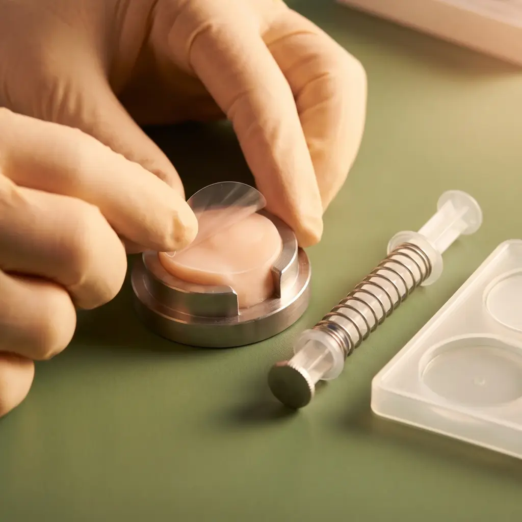
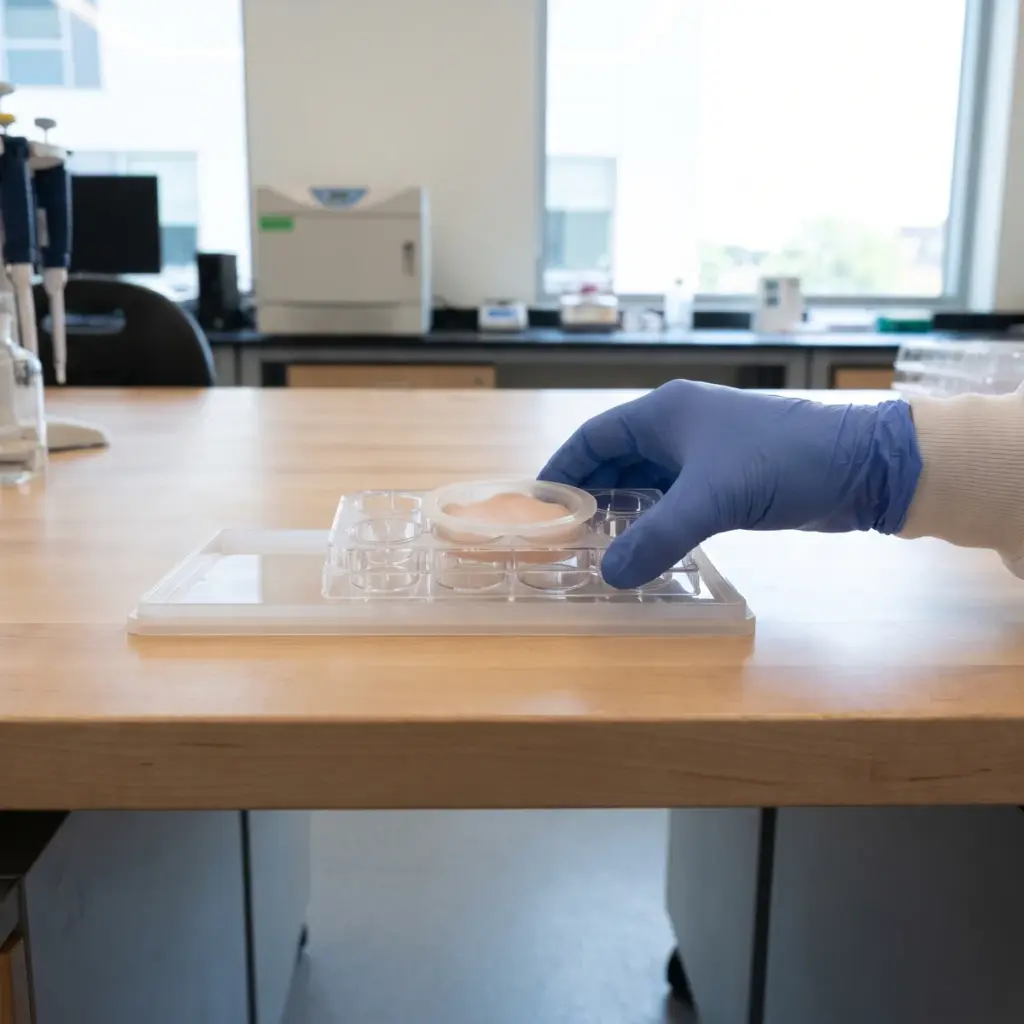
Reconstructed human epidermis shows the full layered structure. After topical or systemic treatment, tissues are fixed and sectioned. Haematoxylin and eosin staining shows layer number and stratum corneum thickness, and immunostaining shows where each marker sits. A granular layer that is better defined, or a marker that extends correctly into the upper layers, is visible directly on the section.
- Early and late differentiation marker expression
- Marker localisation across the epidermal layers
- Epidermal and stratum corneum thickness
- Cornified envelope formation
- Tissue viability controls
Linking maturation to barrier performance
Electrical resistance or tracer permeability can be added to the same tissues to show whether the structural change goes with a tighter barrier. Calcium-dependent signalling is often explored next in calcium gradient maintenance studies.
The report gives quantified marker data with statistics, representative images for each group and a description of the model and dosing.
When to commission keratinocyte differentiation studies
Differentiation data are most useful early, when you have an active candidate and need to know which part of epidermal renewal it affects. A rise in late markers with a thicker, well-organised granular layer supports claims about helping skin build its outer protective layer. A selective effect on filaggrin points you toward filaggrin production testing and a hydration claim.
Choosing between monolayer and 3D designs
Start with monolayer cultures when you have many candidates or need a dose range. They are quicker and give clean transcript and protein data. Move to reconstructed epidermis for the shortlisted active, because histology and marker localisation only make sense in a stratified tissue. Finished formulations, creams in particular, need the 3D model since they are applied to the tissue surface.
Validated differentiation markers and positive controls
Ask which markers the lab has validated antibodies and primers for, and whether it quantifies immunostaining by image analysis or reports images only. Check that it includes a reference active known to promote differentiation, so you can read your result against a positive control. If you plan a barrier claim, ask whether resistance or permeability can be measured on the same tissues.
More in Barrier & Hydration Mechanisms
Barrier & Hydration Mechanisms studies show how an ingredient acts on epidermal lipids, barrier proteins and water-handling pathways in skin models.