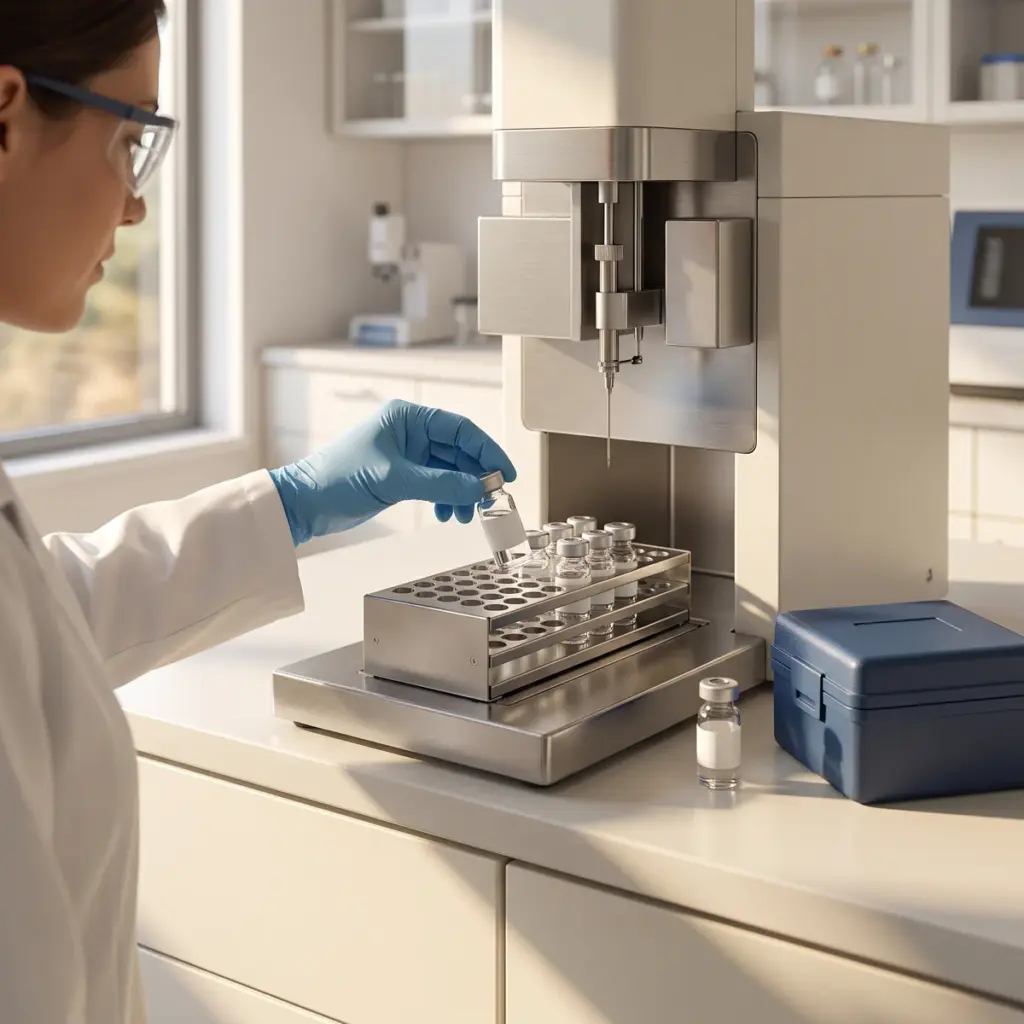
Skin barrier function efficacy testing overview
Do you provide Skin barrier function efficacy testing?
How skin barrier function efficacy testing works
Most of the barrier sits in the stratum corneum, where flattened corneocytes are embedded in ordered layers of ceramides, cholesterol and free fatty acids. Deeper down, tight junctions in the granular layer control what passes between living cells. A barrier study selects the endpoints that match your product’s proposed mechanism and measures them on the same tissue batch.
Reconstructed epidermis and barrier challenge
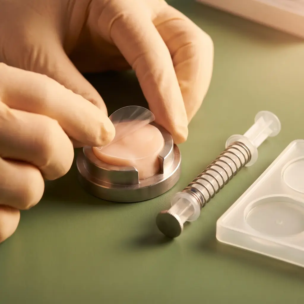
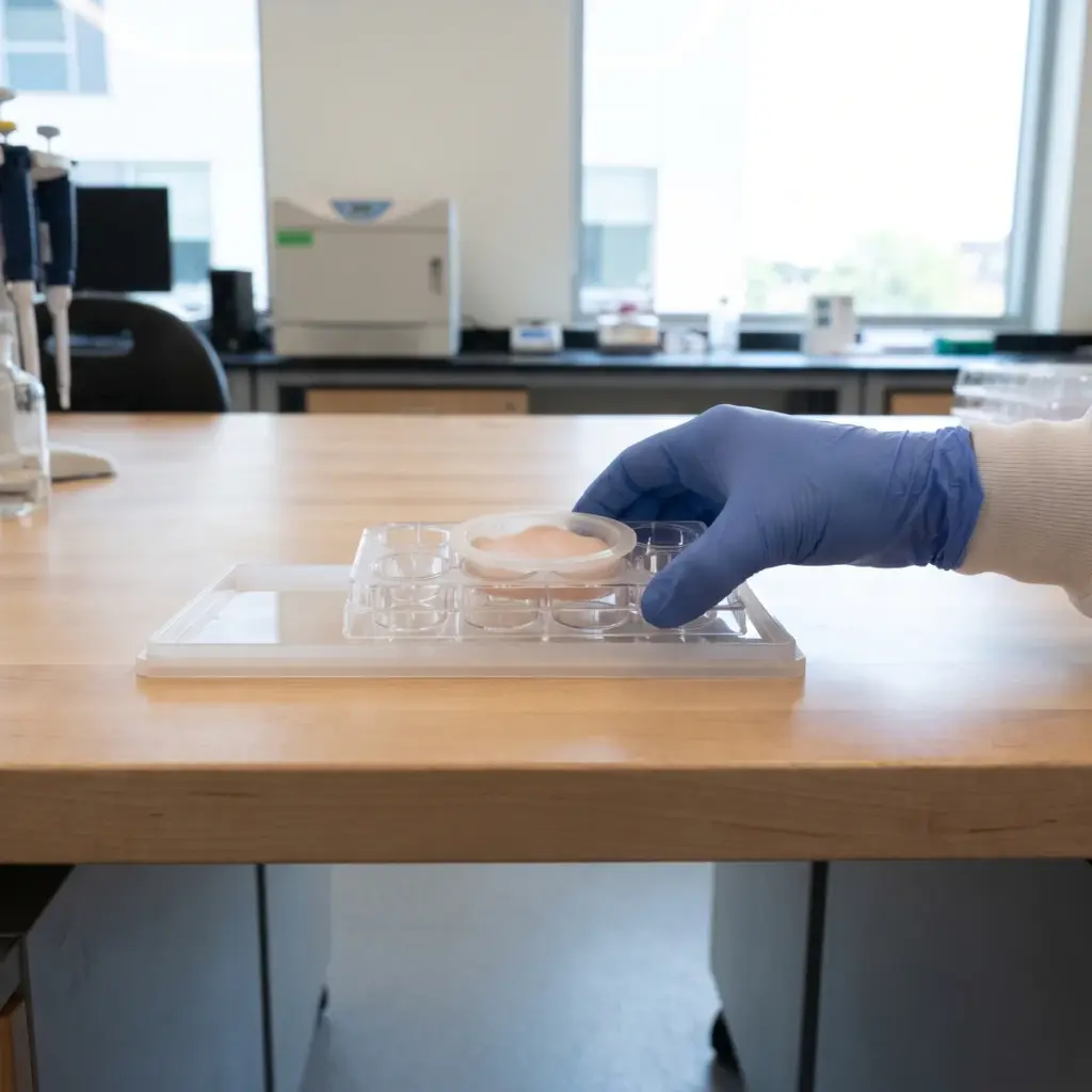
Reconstructed human epidermis is grown at the air-liquid interface, where it stratifies and forms its own stratum corneum. The product is applied to the tissue surface at one or more concentrations. For a repair design, the laboratory first disrupts the barrier with a defined surfactant challenge such as sodium lauryl sulfate, then follows recovery in treated and untreated tissues. For a protection design, the product goes on before the challenge.
Structural and functional barrier endpoints
- Lipid packing and chain order by Raman microspectroscopy
- Ceramide, cholesterol and fatty acid profiles by LC-MS
- Filaggrin, involucrin and loricrin by immunostaining or qPCR
- Tight-junction proteins such as claudin-1 and ZO-1
- Transepithelial electrical resistance and tracer permeability
Tissue viability is measured alongside every treatment. When one mechanism looks promising, it can be studied in depth through ceramide synthesis mechanism studies, filaggrin production assays or tight-junction protein analysis.
Histology, immunofluorescence and barrier marker data reported
The final report describes the model, concentrations, exposure times, controls and statistical tests. It includes histology and immunofluorescence images, marker quantification and functional values for each group, all expressed against untreated and vehicle tissues. From it you can state which barrier mechanisms responded in the model and by how much.
Using skin barrier function efficacy testing in development and claims
Barrier data are used at two points in a project: early, to pick the active or formula worth taking further, and later, to explain a clinical result.
Screening actives before a volunteer study
A barrier panel lets you compare several actives or use levels on one tissue batch before you commit to a clinical protocol. The candidate that restores electrical resistance and lipid packing after the surfactant challenge is the one to carry into a human study measuring transepidermal water loss and skin hydration. The in vitro data then explain the mechanism behind the clinical result.
Wording a barrier mechanism claim
Ingredient suppliers put the results in technical files and marketing literature to support statements such as “supports barrier lipid organisation in a reconstructed epidermis model”. Brands cite them next to human data on the finished product. Build the claim around the endpoint that moved, whether that was lipid packing, differentiation markers or junction proteins.
Preparing your quote request
Send the ingredient or formula, the concentration range, the vehicle and the claim you want to support. Say whether you need a repair design or a protection design. Mention early if the product is strongly coloured or very oily, as residues can interfere with Raman and fluorescence readouts and the laboratory may need to adjust rinsing or sampling.
More in Barrier & Hydration Mechanisms
Barrier & Hydration Mechanisms studies show how an ingredient acts on epidermal lipids, barrier proteins and water-handling pathways in skin models.