Keratinocyte proliferation overview
Do you provide Keratinocyte proliferation?
How keratinocyte proliferation is measured
The epidermis renews itself from a basal layer of dividing keratinocytes. Daughter cells move upwards, differentiate and are finally shed from the surface. This service measures the first step, cell division in the basal population, and can be extended with differentiation markers when you need the wider picture.
Culture and exposure
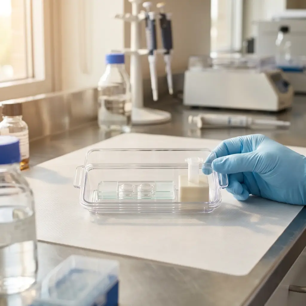
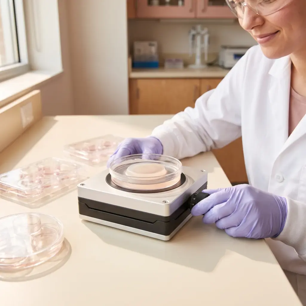
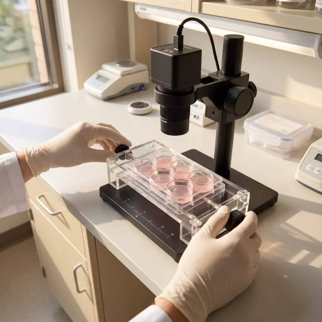
Normal human epidermal keratinocytes are grown in low-calcium medium, which keeps them in a proliferative state, and treated with the test material across several concentrations. Exposure times are chosen to cover at least one division cycle, and untreated, vehicle and growth factor reference wells are included on each plate.
Proliferation endpoints
- BrdU or EdU incorporation into newly synthesised DNA
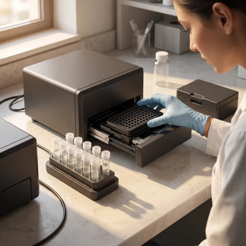
- Cell counts by automated imaging
- Cell-cycle distribution by flow cytometry
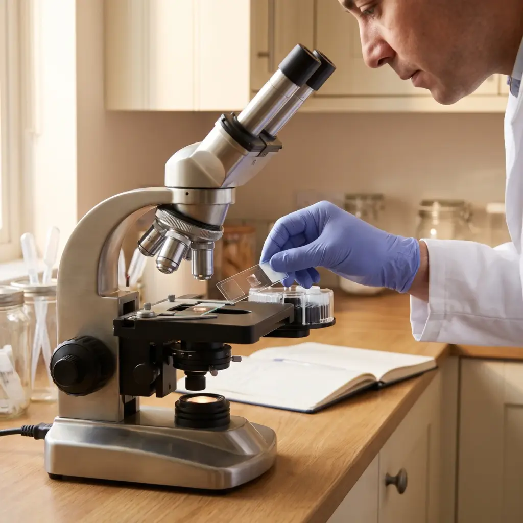
- Ki-67 staining of cycling cells
- Viability by a membrane integrity or metabolic assay
Direct DNA synthesis and cell-count data separate genuine division from a change in metabolic rate, which is why they are preferred over MTT for this question.
Differentiation and tissue context
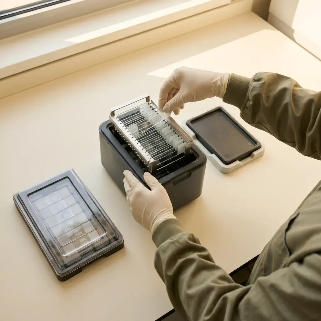
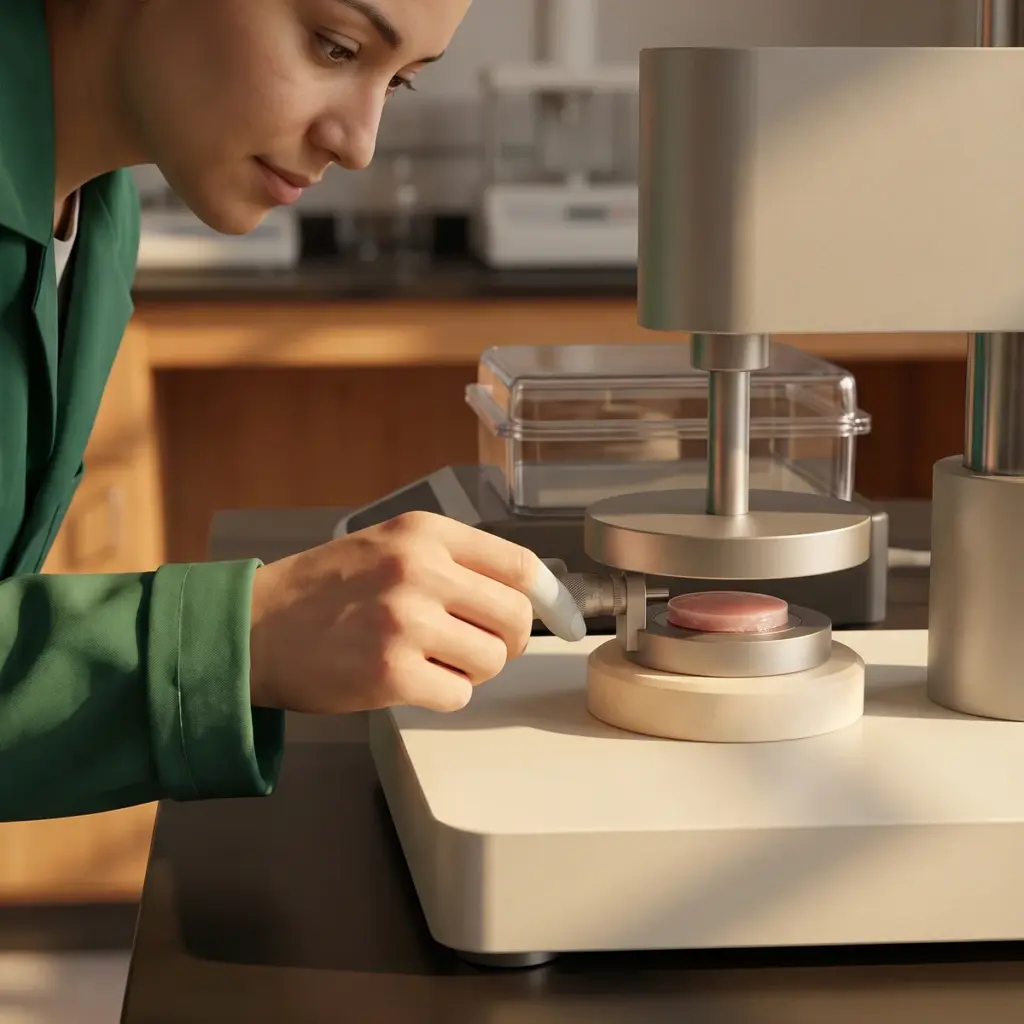
Markers such as keratin 10, involucrin and filaggrin show whether dividing cells go on to differentiate normally. Reconstructed human epidermis lets the laboratory examine layer thickness, basal cell density and tissue organisation after topical application of the finished formula. Results in this format suit a renewal claim that runs from basal division to a well-formed surface.
Proliferation study report
You receive a report with methods, concentration-response data per endpoint, statistical comparison with controls and representative images, plus a recommended concentration range for further work.
Using keratinocyte proliferation data for renewal claims
Brands use proliferation results to explain how an active supports the epidermis from the basal layer upwards.
Where the test fits in a renewal programme
Proliferation data are the cellular starting point for claims about skin renewal and radiance. They work best paired with a dermal study, since many anti-ageing products aim to act on both layers. Pairing this assay with fibroblast activation testing gives a two-layer profile from one laboratory, and the combined results slot into a wider anti-ageing test programme.
Exfoliating acids and enzymes act mainly on shedding at the surface, so their renewal claim is better supported with reconstructed epidermis or volunteer measurements. Actives such as growth factor mimics, peptides and certain vitamins act on the basal layer and are the typical candidates for this test.
Composition, fluorescence and claim details to send
Provide the material’s composition, solvent compatibility and any known colour or fluorescence, since some extracts interfere with imaging readouts. State the claim you have in mind and whether you need differentiation markers in the same study. If you plan to test a finished cream, ask early about reconstructed epidermis, where emulsions are applied topically to the tissue surface.
More in ECM & Skin Ageing
ECM & Skin Ageing testing measures how actives affect collagen, elastin, matrix enzymes, glycation and senescence in skin cell and tissue models.