Depigmentation testing overview
Do you provide Depigmentation testing?
Depigmentation testing in cells and pigmented epidermis
Depigmentation testing looks at the end result, the amount of melanin left after treatment, whatever the route that produced it. It suits products that combine several brightening actives, because the readout captures their joint effect on pigment.
Melanocyte screening
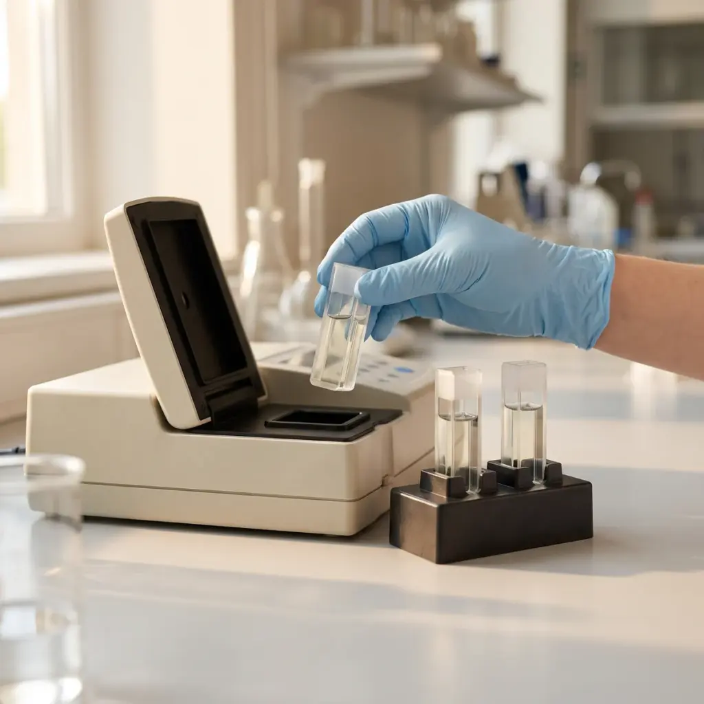
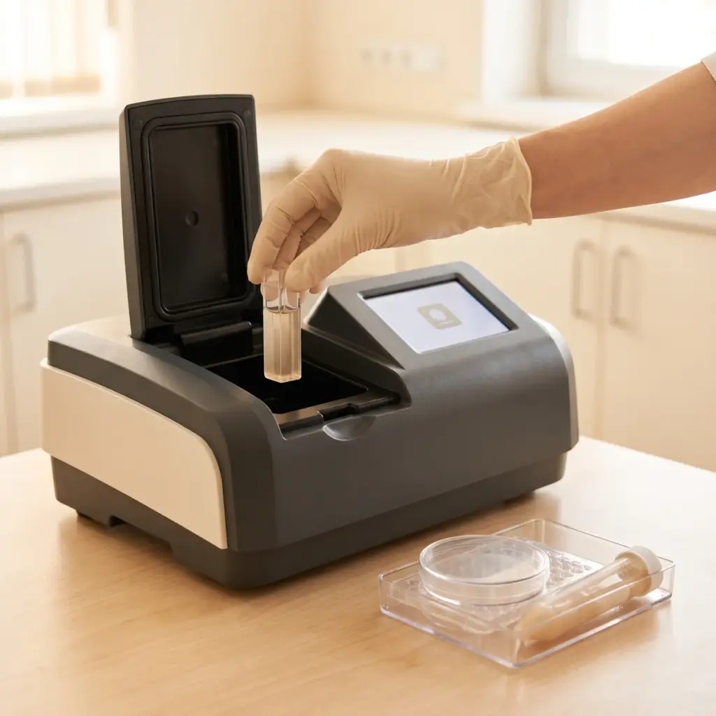
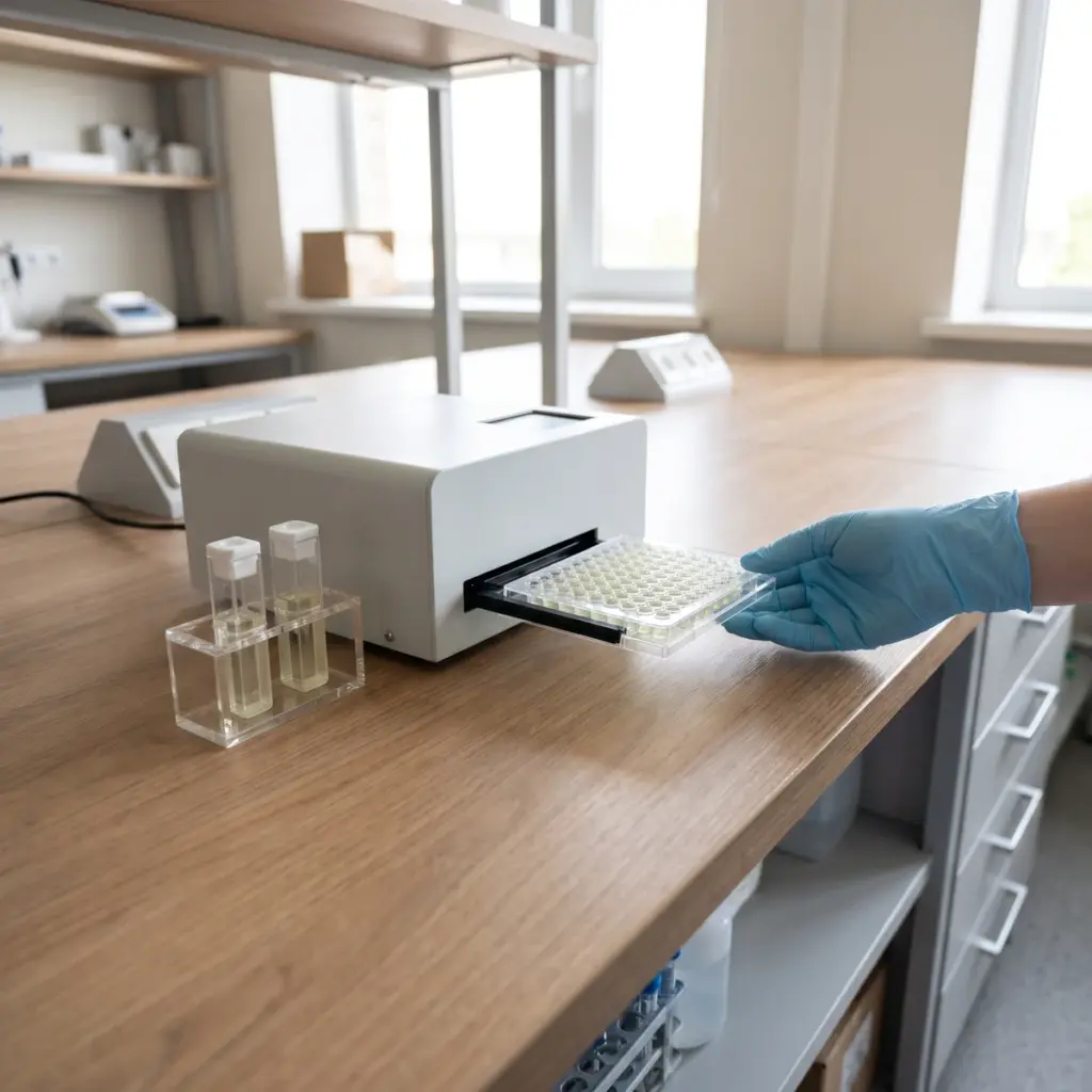
Cultured melanocytes are exposed to the samples across a concentration range. Melanin is extracted and read by absorbance, then normalised to viable cell number or protein. Cellular tyrosinase activity and pigment-gene expression can be added to show how the reduction arose.
Pigmented tissue models
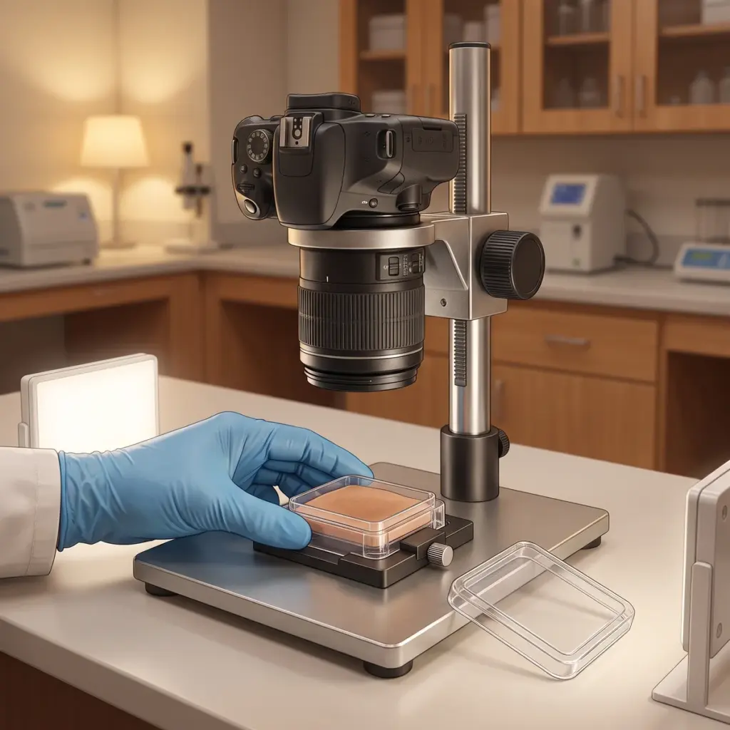
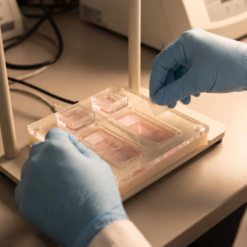
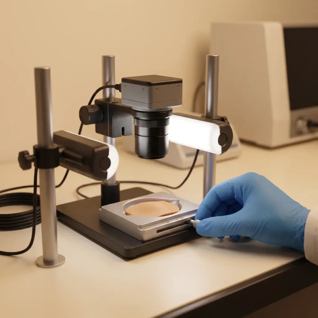
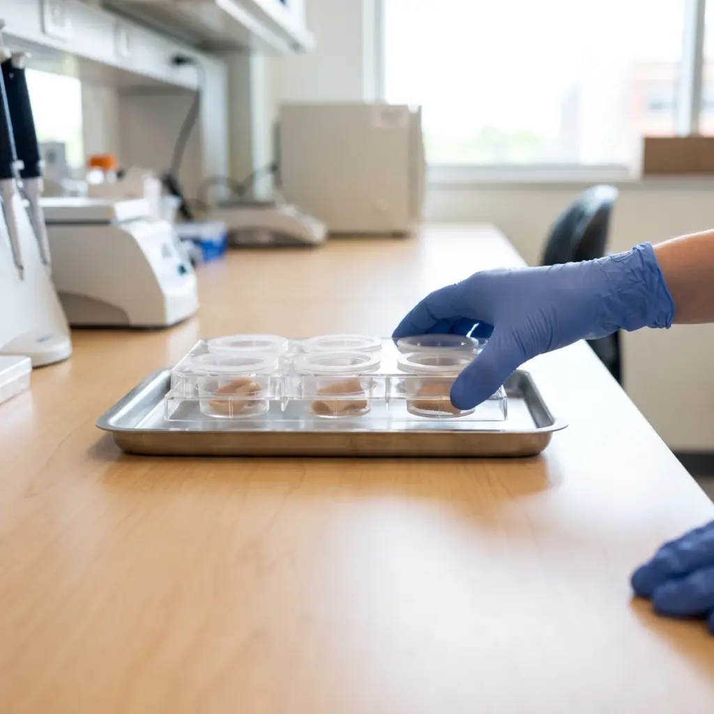
Reconstructed pigmented epidermis accepts topical application, so creams and serums can be tested in their final form. The laboratory reads tissue colour, stains melanin in sections and checks tissue integrity. This format shows pigment distribution through the epidermal layers. Fontana-Masson staining is a common choice for visualising melanin in tissue sections.
Endpoints and deliverables
- Melanin per viable cell or per tissue
- Tissue colour readings and melanin staining
- Concentration-response and time-course data
- Vehicle and reference controls
- Viability and sample interference checks
The report gives melanin values with statistics against controls, tissue images and viability for every condition, so a genuine pigment reduction stands clear of any cell damage. A formula that performs well here is ready for clinical work on volunteers, where colorimetry, melanin index and standardised photography measure visible change in use.
Applying depigmentation results to formula decisions
Order this study when you need to pick a lead brightening formula, compare a new active with a benchmark, or confirm that an active keeps its effect in the finished base. The data supports in vitro statements on melanin reduction and builds the case for a human study.
Preparing samples and a brief
Send raw materials with their solvents and test concentrations, and finished products in their final packaging form. Name the benchmark you want to beat. If your claim depends on a mechanism, mention it, so the laboratory can add a tyrosinase inhibition screen or gene expression panel.
Cell and pigmented tissue capability to check
Choose a provider that can test both raw materials in cells and formulas on pigmented tissue, so results stay comparable as the project moves forward. Ask how it corrects for coloured samples and how it reports viability. Laboratories with links to clinical partners make the move from tissue data to the wider pigmentation efficacy programme straightforward.
More in Pigmentation & Melanogenesis
Pigmentation & Melanogenesis testing studies how actives change melanin synthesis, melanocyte behaviour and pigment transfer in lab models.