Pigment vesicle dynamics overview
Do you provide Pigment vesicle dynamics?
Melanosome trafficking and pigment vesicle imaging
Melanosomes pass through four maturation stages, from unpigmented early vesicles to fully melanised organelles. Mature melanosomes are carried along microtubules and then actin filaments to the tips of the dendrites, ready for transfer. Pigment vesicle dynamics testing measures where an ingredient changes this route, whether by slowing maturation, holding organelles back or altering their spread.
Labelling and microscopy
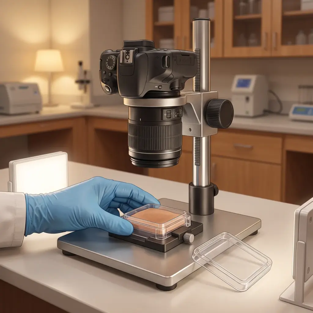
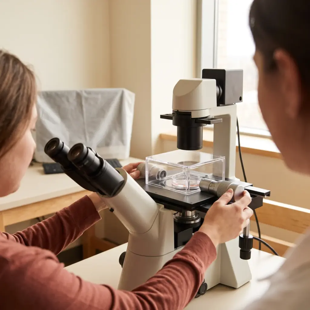
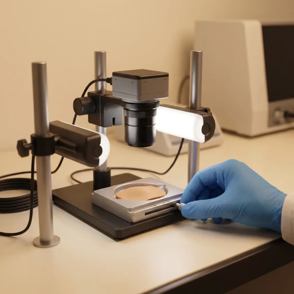
Melanocytes are treated with the test sample and controls, then stained with antibodies against melanosome proteins or imaged by brightfield for pigmented organelles. Confocal imaging gives the position of each vesicle. Live-cell time-lapse, where the model permits, records movement speed and direction. Image analysis software then assigns each vesicle to a cell region and a maturation class.
Trafficking parameters
- Melanosome count per cell
- Proportion of early and mature organelles
- Distribution between cell body and dendrites
- Movement tracks and perinuclear clustering
- Viability and label controls
Reading trafficking beside synthesis
Melanin content and tyrosinase activity describe how much pigment is made. Vesicle data describe where it goes. Measuring both lets the laboratory say whether an ingredient acts on formation, on transport or on both. Co-culture with keratinocytes extends the study to the final hand-over, and pigment transfer inhibition testing quantifies that step directly.
Deliverables include annotated images and time-lapse files where recorded, quantitative tables per condition and a short mechanistic interpretation.
Using melanosome dynamics in an ingredient dossier
Commission this study when your active has a proposed transport mechanism, or when it lowers visible pigment in tissue models without much change in melanin synthesis. Trafficking data gives clear, image-led evidence for technical presentations and supplier files. It also helps explain why two actives with similar melanin results perform differently in tissue.
Details to share with the laboratory
Describe the mechanism you expect, for example clustering near the nucleus or reduced movement into dendrites. Supply the sample, solvent and a concentration range that keeps melanocytes healthy. Say whether you need live-cell recordings, because they need specific equipment and labelling methods.
Choosing an imaging partner
Ask which melanosome markers the provider uses and how it confirms that labelling leaves cell behaviour unchanged. Check how many cells are tracked and whether analysis is automated. Request a sample image set from a previous study to judge resolution and labelling quality. A provider that can also measure dendritic process reduction in the same images gives you transport and morphology data from one experiment.
More in Pigmentation & Melanogenesis
Pigmentation & Melanogenesis testing studies how actives change melanin synthesis, melanocyte behaviour and pigment transfer in lab models.