Dendritic process reduction overview
Do you provide Dendritic process reduction?
Quantifying melanocyte dendricity by image analysis
Each melanocyte contacts many keratinocytes through its dendrites. Longer and more branched dendrites give the cell more contact points for pigment delivery. A reduction in dendricity is a measurable structural endpoint, and it gives a brightening active a mechanism that sits downstream of melanin synthesis.
Culture and imaging
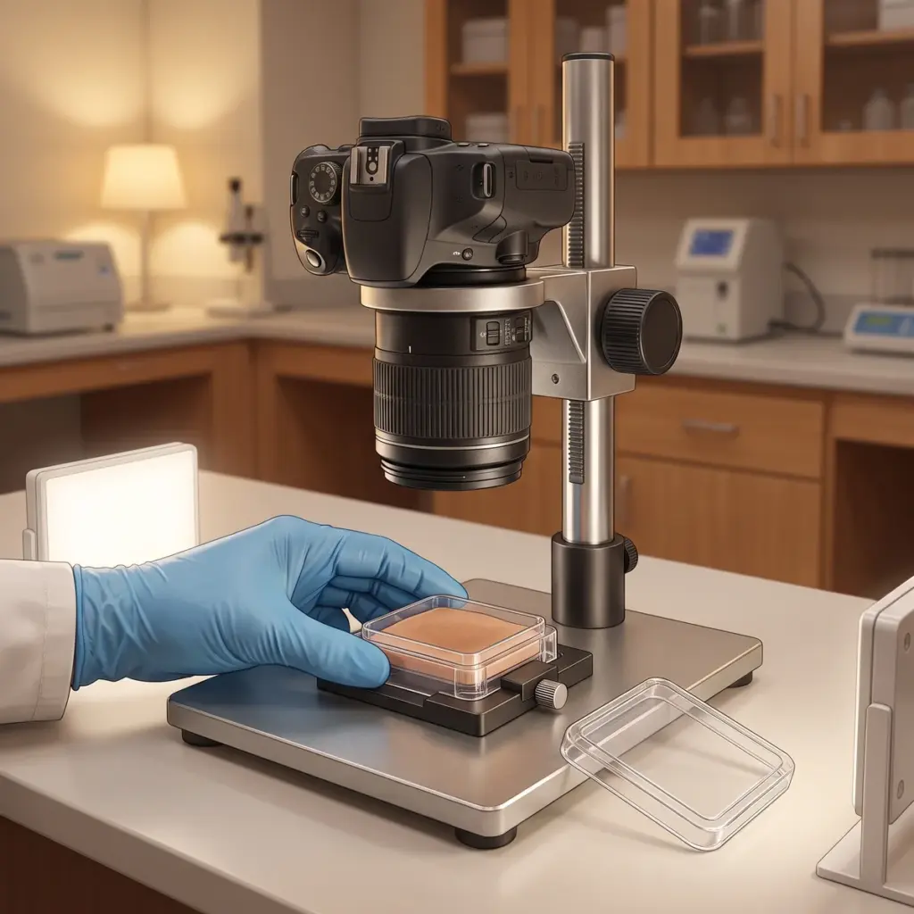
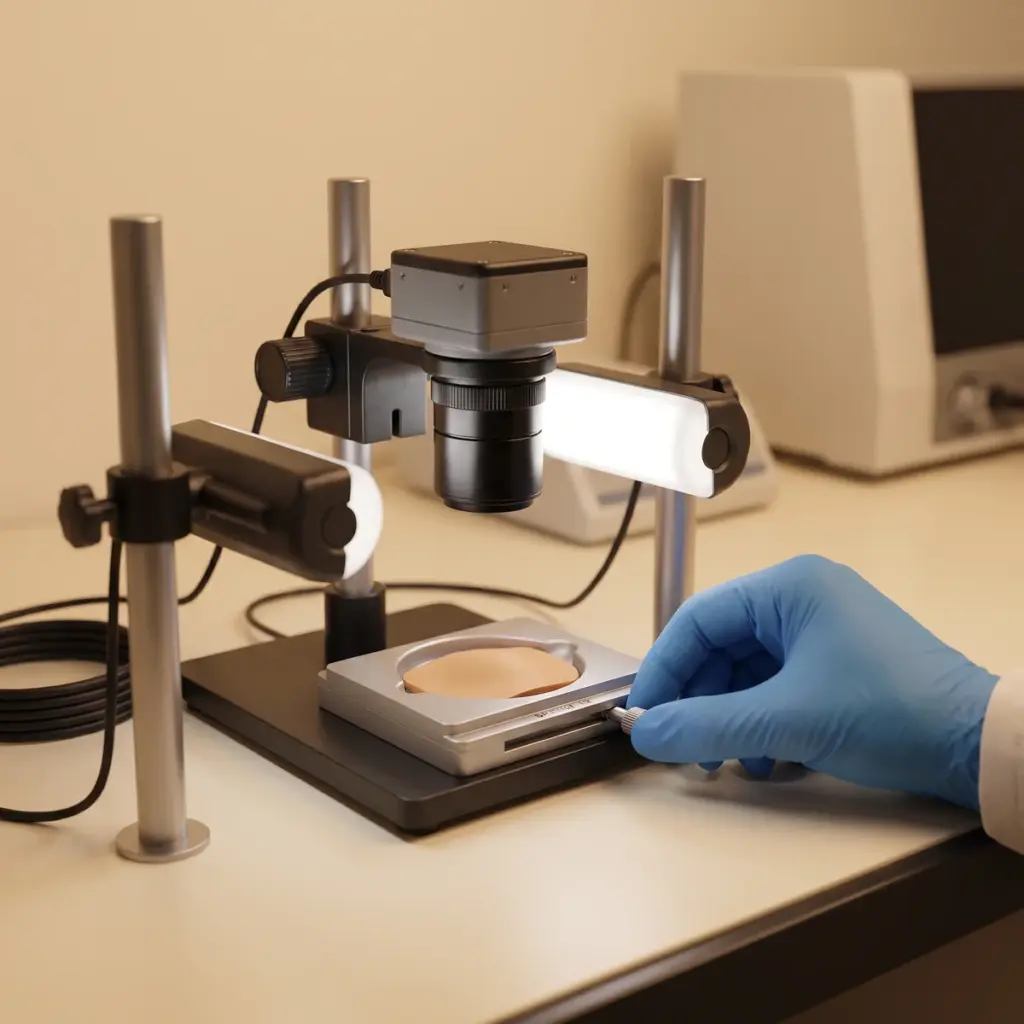
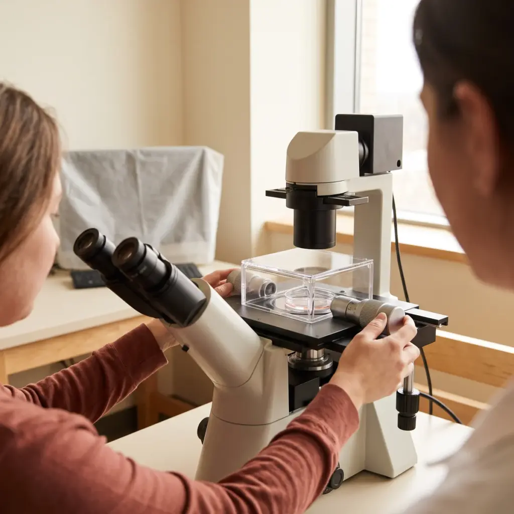
Human melanocytes are seeded at a density that leaves room for dendrites to spread. After treatment with the test sample and controls, cells are fixed and stained, often for cytoskeletal proteins, then imaged on a microscope at fixed settings. Co-culture with keratinocytes can be used when cell contacts matter. Image fields are chosen at random or by a fixed grid, so the cells analysed represent the whole well.
Morphology parameters
- Dendrites per cell and mean dendrite length
- Branch points and total dendritic area
- Cell body shape and spread
- Cell attachment and viability at each concentration
Mechanism add-ons
Rac1 and CDC42 are small GTPases that drive dendrite extension. Measuring them, where the study calls for it, can explain why a sample changes cell shape. Viability and attachment data separate a specific morphology effect from cells that are simply stressed or detaching. The report includes representative images, the measurement definitions used and statistics against controls. To show what happens to pigment delivery itself, the natural next study is pigment transfer inhibition.
Planning a dendritic process reduction study
This study fits actives proposed to change pigment distribution, such as ingredients thought to act on the cytoskeleton. It is also useful when a brightening ingredient shows little effect on tyrosinase and you need another mechanism to explain its performance.
Co-culture format and Rac1 or CDC42 choices
Supply the sample, its solvent and a concentration range you expect to be well tolerated by melanocytes. State whether you want monoculture or keratinocyte co-culture, and whether Rac1 or CDC42 measurements are needed. If you hold earlier melanin or enzyme data, share it so the laboratory can choose matching concentrations and read the morphology result against your existing pigment findings.
Judging imaging providers
Ask how the laboratory defines a dendrite and a branch point, and whether image analysis is automated or blinded. Check how many cells are measured per condition and whether you will receive raw images. Providers that also run melanosome tracking studies can follow pigment along the dendrites in the same cell model.
More in Pigmentation & Melanogenesis
Pigmentation & Melanogenesis testing studies how actives change melanin synthesis, melanocyte behaviour and pigment transfer in lab models.