Pigment transfer inhibition overview
Do you provide Pigment transfer inhibition?
Measuring melanin transfer to keratinocytes
Pigment that colours the skin sits mostly in keratinocytes, which receive it from melanocytes. Slowing that hand-over leaves less pigment in the upper epidermis. This service quantifies transfer as its own endpoint, so a transfer effect can be told apart from reduced melanin synthesis.
Co-culture and tissue models
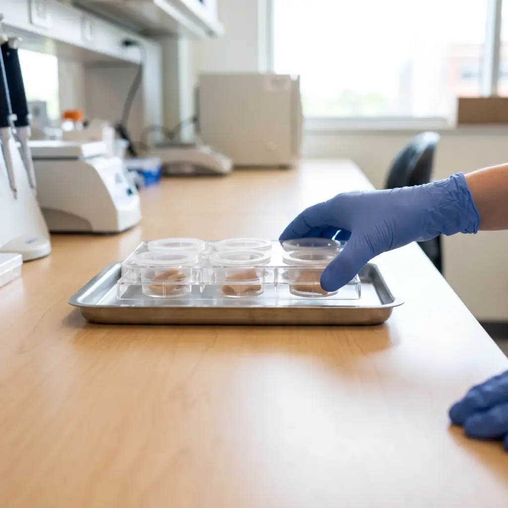
Human melanocytes and keratinocytes are grown together at a defined ratio, or a pigmented reconstructed epidermis is used for a skin-like structure. The test sample is applied in medium or topically. Cell-specific markers let image analysis or flow cytometry assign pigment to each cell type.
Endpoints in a transfer study
- Pigment content in identified keratinocytes
- Melanin content and cell number in donor melanocytes
- Viability of both cell populations
- Concentration-response with transfer-relevant controls
Controls and supporting measures
A synthesis inhibitor such as kojic acid or arbutin lowers total pigment, which makes it a useful contrast to a transfer-specific effect. The laboratory chooses controls that match the mechanism under test. Dendrite length, branching and cell contacts can be measured in the same cultures to explain a change in delivery. Reporting donor pigment beside recipient pigment shows whether lower keratinocyte pigment reflects slower transfer or less melanin to begin with.
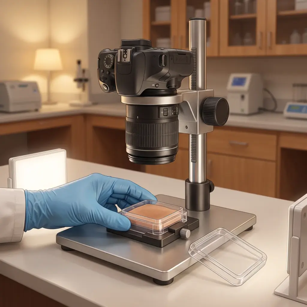
The report includes images, quantitative data for each cell type and statistics against controls. Flow cytometry data, where used, are supplied with the gating strategy.
Commissioning a pigment transfer inhibition study
This study suits actives proposed to work at the melanocyte-keratinocyte interface, and it adds a second mechanism to ingredients that also lower melanin synthesis. The results support a mechanism statement on brightening or even-tone ingredients. Transfer data is also persuasive in technical sales material, because images of pigment-free keratinocytes make the mechanism easy to see.
Sample, concentrations and prior melanin data to send
Provide the sample, its solvent and the concentrations you want tested, plus any earlier melanin or dendrite morphology data. Say whether the sample will be applied in medium or topically to a tissue model, since a formula usually calls for the tissue format.
Questions for prospective providers
Ask how the laboratory identifies recipient cells and assigns pigment to them. Check which positive control it uses for transfer and how it reports donor melanin. Ask too for the melanocyte to keratinocyte ratio, since it changes how much pigment each recipient cell receives. A provider that also offers pigment vesicle dynamics can show the full route from melanosome transport to keratinocyte uptake.
More in Pigmentation & Melanogenesis
Pigmentation & Melanogenesis testing studies how actives change melanin synthesis, melanocyte behaviour and pigment transfer in lab models.