Growth-factor stimulation


Growth-factor stimulation overview
Who provides Growth-factor stimulation
Companies providing Growth-factor stimulation
Growth-factor stimulation assays: models, markers and readouts
The dermal papilla at the base of the follicle sends signals to the surrounding epithelial cells that influence growth phase and fibre production. Several of these signals are growth factors. VEGF relates to the blood supply around the follicle, IGF-1 to cell proliferation and survival, and FGF-7 (also called keratinocyte growth factor) to the epithelial cells that build the shaft. The study selects the factors that fit the mechanism you want to describe.
Dermal papilla cells, keratinocytes and co-cultures
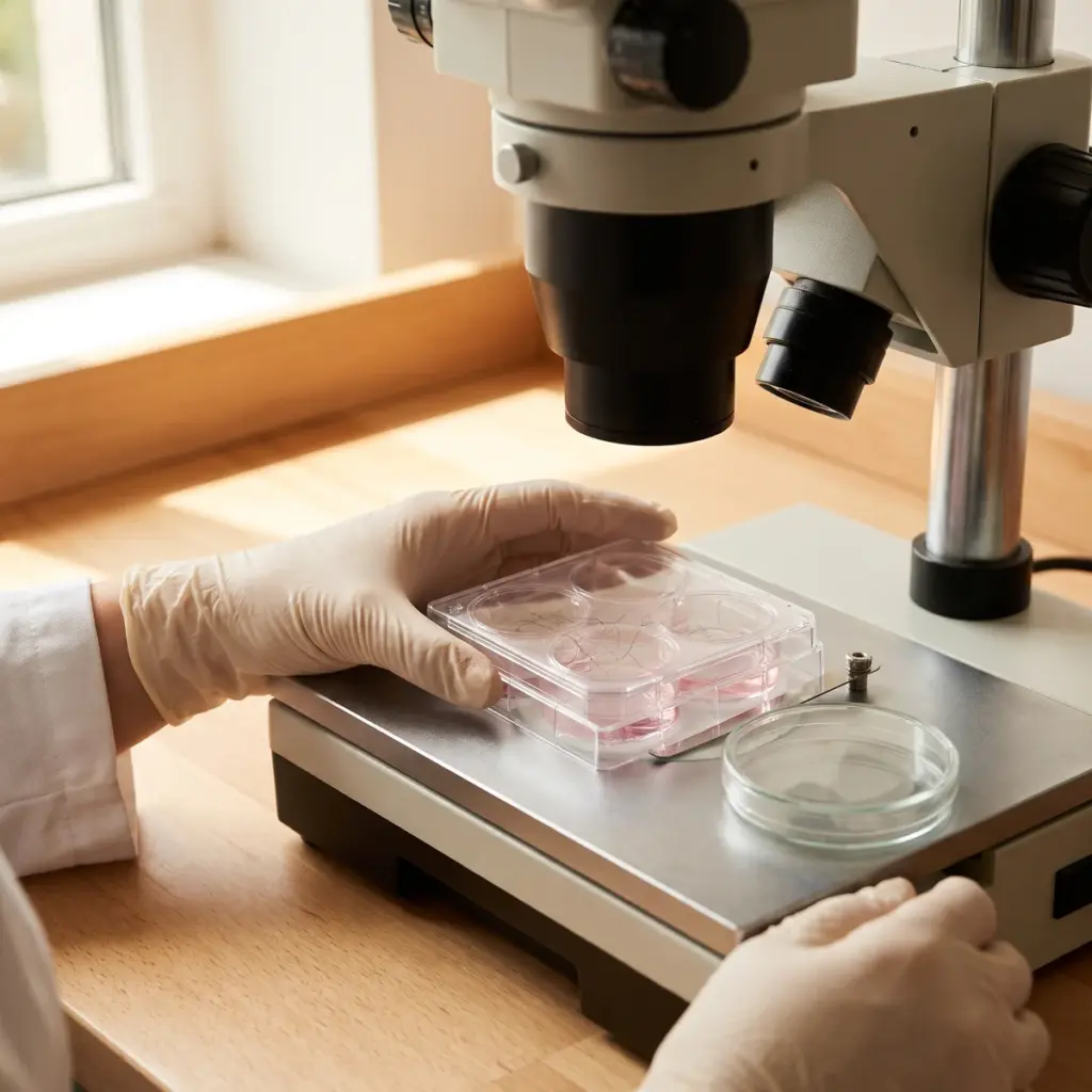
Primary human dermal papilla cells are the most common model. Follicular keratinocytes are used when the target factor is produced by the epithelium, and co-cultures of both cell types show paracrine effects between them.
Gene and protein measurement
Quantitative RT-PCR measures transcript levels shortly after treatment. ELISA or multiplex immunoassays measure the protein secreted into the culture medium over a longer window. The two readouts answer different questions, so a good report presents both and states each separately.
- Transcript fold change against vehicle control
- Secreted protein concentration in conditioned medium
- Concentration-response and time-course profiles
- Viability and cell number for normalisation
- Reference stimulant as positive control
Growth-factor tables, graphs and follicle interpretation
You receive the protocol, the identity of the cells and donors, data tables and graphs for each factor and time point, statistics against controls, and a short interpretation linking the profile to follicle biology.
Where growth-factor data fits in hair active development
Growth-factor results are usually generated early, while the formula is still open and several actives are in contention.
Screening and positioning an ingredient
Growth-factor panels are a practical screening tool. Several extracts or concentrations can be compared in one run, and the most active candidate goes forward. The resulting profile also gives marketing a concrete mechanism, such as “stimulates VEGF release by dermal papilla cells in vitro”, for the ingredient data sheet.
Combining it with other follicle studies
Growth-factor release is one part of the signalling picture. Pairing it with stem cell activation data shows how the signal reaches the cells that respond to it. Whole-follicle culture then tests the effect on shaft elongation, and a clinical study on the finished scalp product measures hair density or growth rate on volunteers.
Target factors, solvent and donor details to provide
Tell the laboratory which factors you are interested in and why, the concentration range you plan to use in formulation and the solvent your ingredient needs. Coloured or turbid extracts can interfere with plate-reader assays, so mention them. Ask whether the lab measures secreted protein as well as gene expression and whether it uses dermal papilla cells from more than one donor.
More in Hair Follicle & Fibre
Hair Follicle & Fibre studies test how an active works on the living follicle and the hair shaft: growth signals, androgens, keratin and pigment.