Skin layer distribution overview
Do you provide Skin layer distribution?
Mapping skin layer distribution of cosmetic ingredients
Total permeation tells you how much active crossed the skin. Distribution tells you where the retained part sits, which is often the more relevant figure for a cosmetic active. The study is planned around the compartments that matter for the ingredient, and the method is chosen to separate them cleanly.
Separating the layers
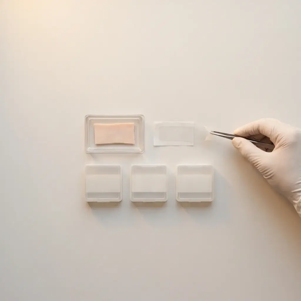
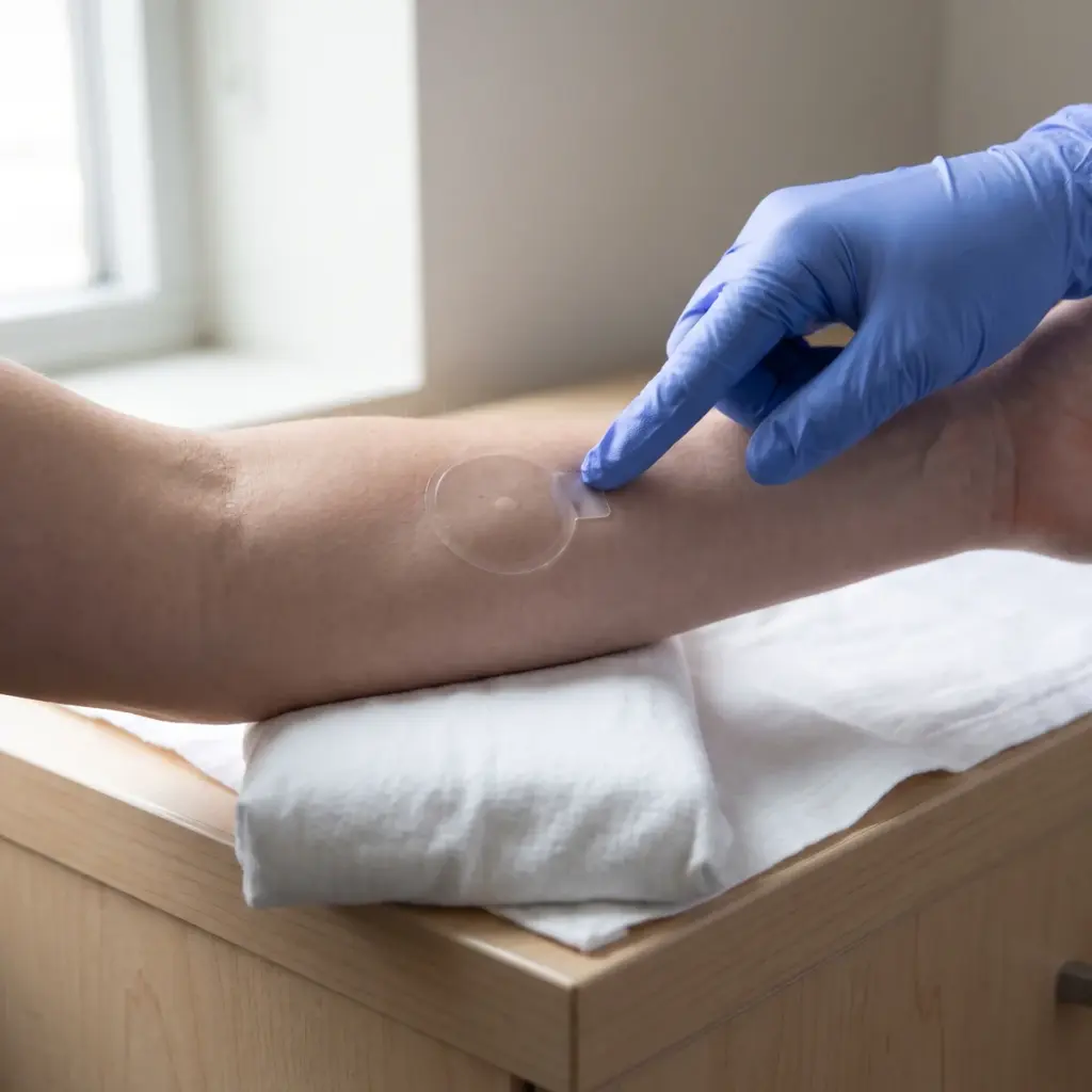
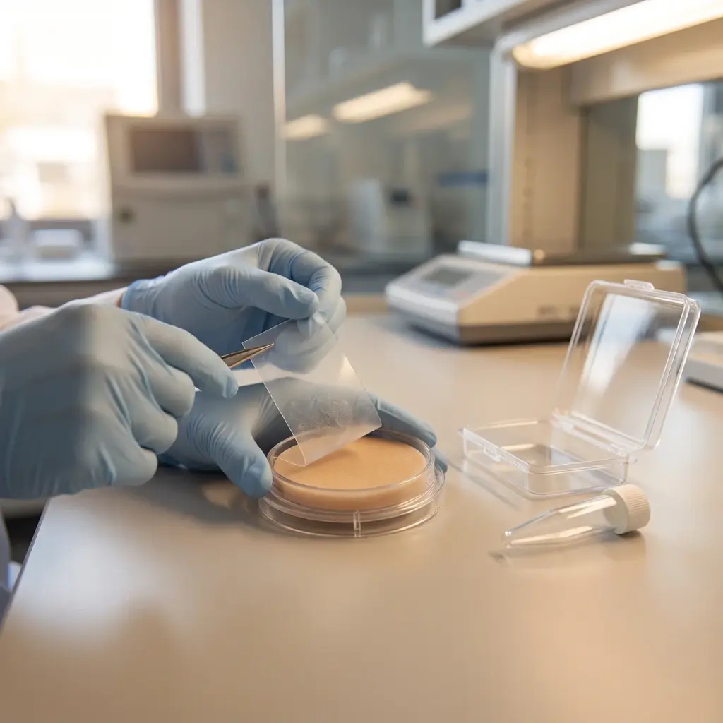
- Tape stripping to collect the stratum corneum
- Heat or enzymatic separation of epidermis from dermis
- Cryosectioning parallel to the surface for fine depth slices
- Solvent extraction of each compartment
- HPLC or mass spectrometry to quantify the active
Before separation, the skin surface is washed so unabsorbed product does not contaminate the deeper fractions. Extraction recovery is determined for each tissue type, because dermis and stratum corneum hold an ingredient differently.
Imaging the active in tissue
Where the ingredient fluoresces, or a fluorescent label stays bound to it, confocal laser scanning microscopy shows its position in cross-sections or optical slices. Raman microspectroscopy can follow some unlabelled molecules by their spectral signature. Imaging gives a visual map, and laboratories usually pair it with extraction data to put numbers on what the images show.
Active per skin layer and depth slice
The report gives the amount of active per compartment or per depth slice, expressed per unit area and as a percentage of the applied dose, alongside surface wash and recovery figures. Images, where used, are annotated with the layer boundaries. Comparing formulas side by side shows which one places more active in the layer you are targeting.
Using skin layer distribution data in formula design
Layer data are most valuable when you know where your active is supposed to work.
Matching the carrier to the target site
Different actives work at different depths. A UV filter or film former should stay on top. A tyrosinase-inhibiting ingredient is aimed at the epidermis, where melanocytes sit. A collagen-signalling peptide is aimed at the dermis. Distribution data let you check whether a liposome, nanoemulsion or other carrier moves the active towards its intended layer, or keeps it at the surface when that is the goal. For a direct comparison of carriers on a fixed objective, an active delivery assessment builds on the same methods.
Combining distribution with permeation
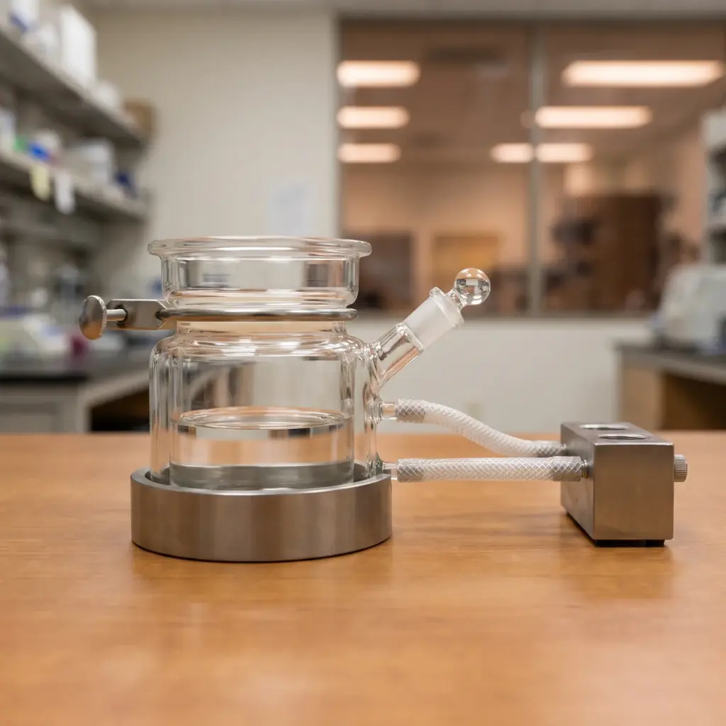
Many projects run distribution and permeation in one experiment. The skin sits in a Franz diffusion cell during exposure, the receptor fluid is sampled for permeation, and the tissue is separated at the end. One set of skin samples then gives you both the amount crossing the skin and its location within it, and the combined data feed into exposure assessment as well as formulation choice.
Layers, sampling times and sectioning method to specify
State which layers matter for your active and at what times you want them sampled. Ask whether the laboratory uses heat separation, cryosectioning or both, and what depth resolution its sectioning gives. If you want imaging, check whether your ingredient can be detected directly or needs a label, and whether the laboratory confirms imaging results by extraction.
More in Skin Absorption Studies
Skin absorption studies measure how much of a cosmetic ingredient is released, enters the skin and reaches each layer after application.