Proteasome activity overview
Do you provide Proteasome activity?
Proteasome activity assays: substrates, stress models and readouts
The proteasome is a barrel-shaped enzyme complex. Its 20S core degrades oxidised proteins directly, while the 26S form, with its regulatory cap, handles proteins tagged with ubiquitin. Testing measures how a cosmetic ingredient changes these activities in skin cells.
The three catalytic activities
Each activity cuts proteins after a different type of amino acid, and each has its own fluorogenic substrate:
- Chymotrypsin-like activity, read with Suc-LLVY-AMC
- Trypsin-like activity, read with Boc-LRR-AMC
- Caspase-like activity, read with Z-LLE-AMC
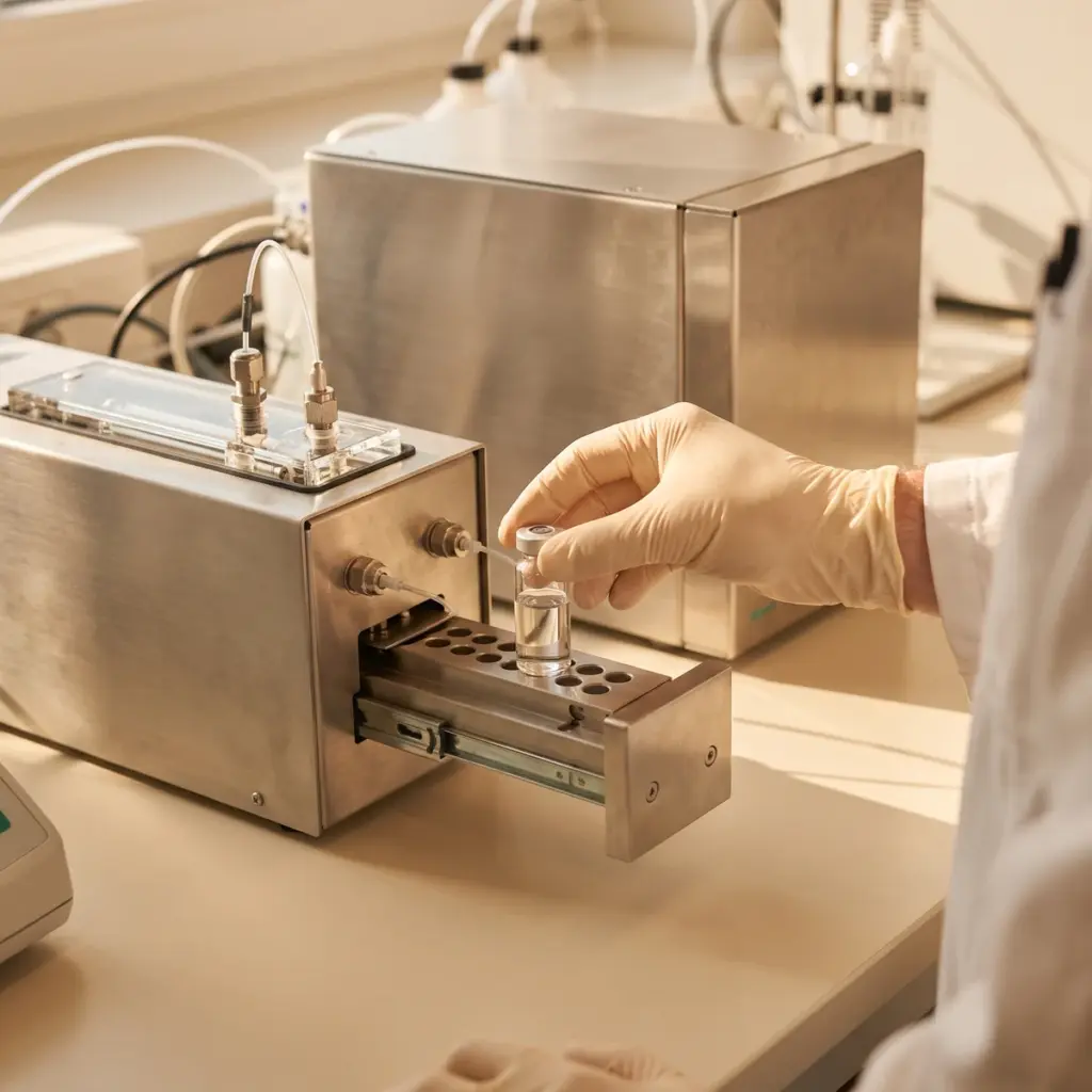
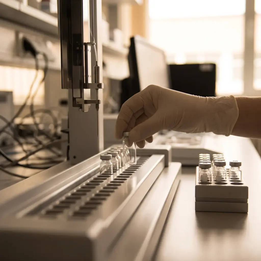
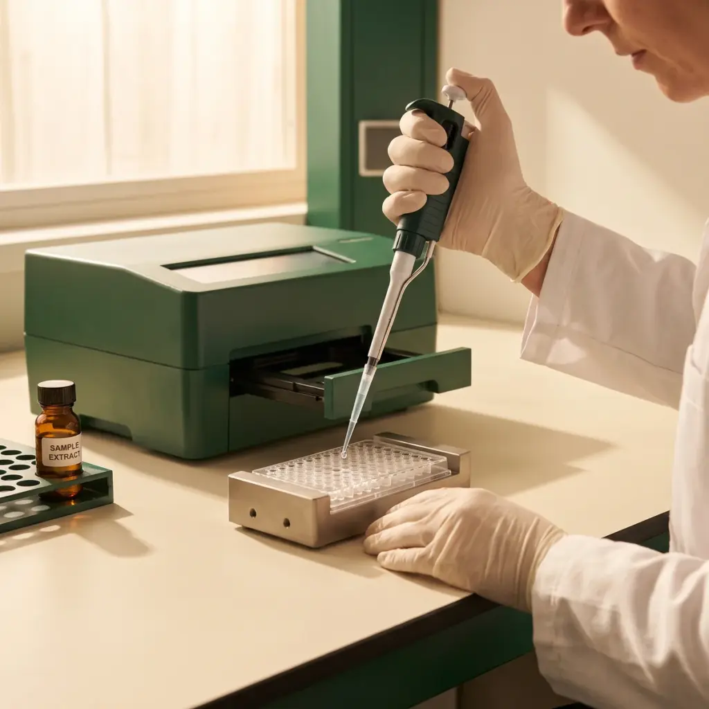
The proteasome cleaves the substrate and releases a fluorescent group, which a plate reader follows over time. Parallel wells with a proteasome inhibitor such as MG132 or epoxomicin give the background, so the reported value is proteasome-specific. Checks for autofluorescence of the test material are run on the same plate.
Stress and recovery design
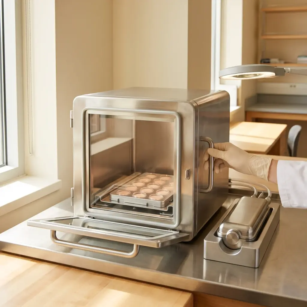
Fibroblasts or keratinocytes are pre-treated with the ingredient, then challenged with hydrogen peroxide, UVA or a defined particulate pollutant. Activity is measured at several points during recovery. Oxidised protein, measured as carbonyl groups, and protein aggregates are quantified on the same samples, along with viability.
Catalytic site activity and recovery time-courses delivered
You receive activity values for each catalytic site normalised to protein content, concentration-response curves, recovery time-courses and the matching protein damage data. Proteasome studies are often run alongside cellular autophagy testing, which handles larger cargo such as protein aggregates and damaged organelles.
Putting proteasome activity results to work in product development
Proteasome data is quick to generate on plates, so brands tend to use it twice: first to pick an active, then to support the protein-clearance wording in the claim file.
Screening actives and setting the dose
The plate-based format makes proteasome activity a practical early screen. You can compare several botanical extracts or peptides on the same plate, then take the most active one into a dose-response study to fix the concentration for your formula. Results from aged donor fibroblasts, or from cells after oxidative challenge, give the most relevant picture for an anti-ageing positioning.
Activity data on its own supports wording about stimulating the cell’s protein clearance system. Paired with lower carbonyl levels, it supports a statement that the active helps skin cells clear oxidised proteins. Brands working on a broader cellular defence claim often add stress response protein data from the same experiment.
Catalytic activities and stress model to specify
Tell the laboratory which catalytic activities you want measured, or whether a first screen on chymotrypsin-like activity is enough. Give the solvent and the solubility of your ingredient, since coloured or fluorescent extracts need extra interference controls. State whether you need a stress model and which challenge fits your marketing claim, whether UV, pollution or general oxidative stress.
More in Xenobiotic & Cellular Clearance Studies
Xenobiotic & Cellular Clearance Studies show how skin cells respond to pollutants and foreign compounds, and how an active supports their recycling system.