Cellular autophagy overview
Do you provide Cellular autophagy?
How cellular autophagy is measured in skin cells
Autophagy is the cell’s own recycling route. A double membrane forms around worn proteins, lipid droplets or damaged mitochondria, the resulting autophagosome fuses with a lysosome, and lysosomal enzymes break the contents down for reuse. The activity of this pathway declines in aged skin cells, which is why it is studied for anti-ageing actives.
Cell models and treatment design
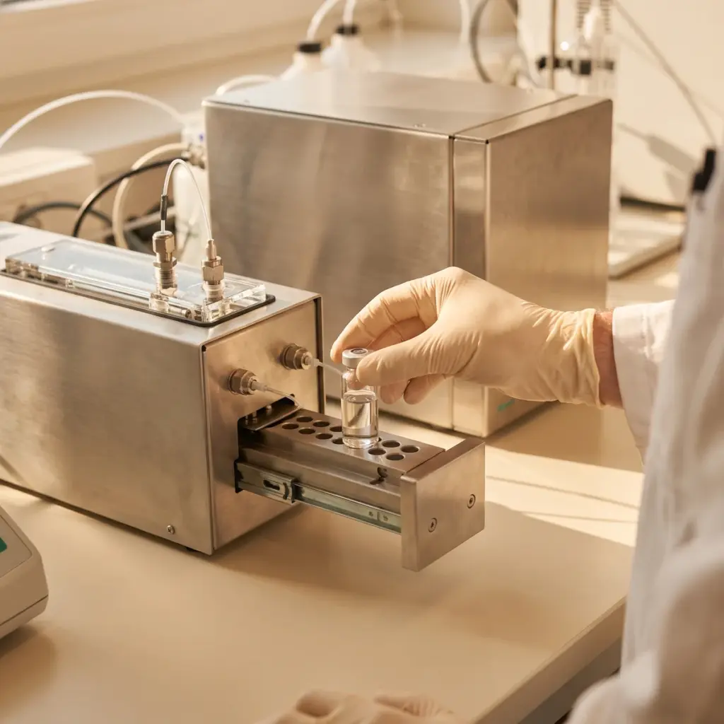
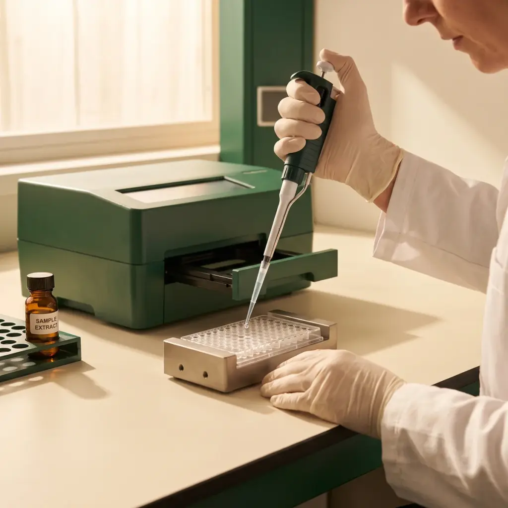
Human keratinocytes or dermal fibroblasts are treated with the test ingredient across a concentration range and over several timepoints. Studies can run at baseline or under a stress such as UV exposure, oxidative challenge or a particulate pollutant. A known autophagy inducer, for example rapamycin or nutrient starvation, gives a positive control.
Autophagy markers and flux
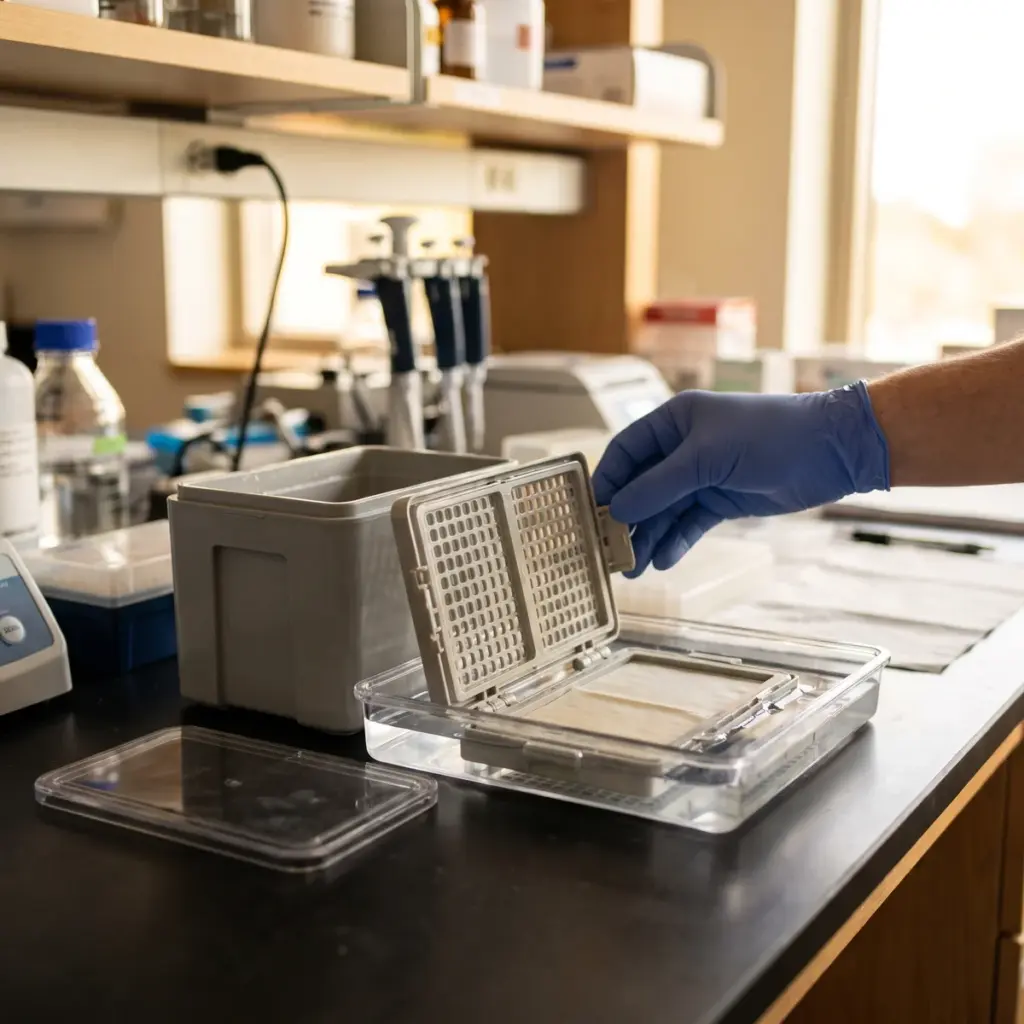
- LC3-II levels by western blot
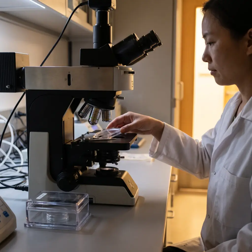
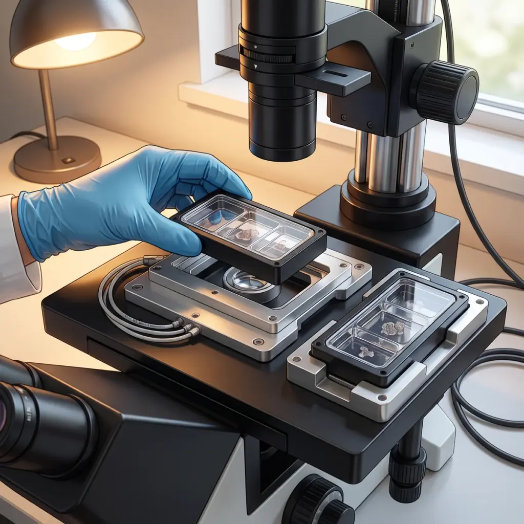
- LC3 puncta counted by fluorescence microscopy
- p62 protein levels
- Lysosomal and cathepsin activity
- Mitophagy markers where mitochondrial quality is part of the claim
Flux is the measurement that matters most. The laboratory repeats the marker readings with and without a lysosomal inhibitor such as bafilomycin A1 or chloroquine. If LC3-II builds up further when degradation is blocked, the ingredient has increased the rate at which material passes through the pathway.
Autophagy markers, puncta counts and flux data reported
The report gives concentration-response and time-course data for each marker, representative images with quantified puncta per cell, flux calculations and viability. It names the stage of the pathway each readout describes. Many brands pair this with proteasome activity testing, since the proteasome is the other main protein clearance route in the cell.
Building a claim on cellular autophagy data
Autophagy results are most useful early, while you are still choosing the active and its dose, and later as the mechanism chapter of an anti-ageing or anti-pollution claim file.
Claims autophagy results can support
A demonstrated increase in autophagic flux supports in vitro mechanism wording such as an active that stimulates cellular recycling or the self-cleaning capacity of skin cells. Under a pollution or UV challenge, the same readouts support a statement that the active helps cells maintain their recycling activity under stress. These statements usually sit in the mechanism section of a claim dossier, ahead of instrumental or clinical results on the finished product.
Combining autophagy with damage endpoints
Autophagy data becomes more persuasive when it sits next to a measure of what the cell cleared. Oxidised protein, lipid peroxidation or mitochondrial function readouts in the same experiment link the higher flux to a cleaner cell. Stress response protein studies add heat shock protein and antioxidant response data on the same model.
Selecting an autophagy laboratory
Ask whether the flux protocol with a lysosomal inhibitor is part of the standard design or an extra. Check that image analysis is automated and counts puncta across many cells per condition, and that the laboratory runs its own positive control in every experiment. Prior work on primary human skin cells is a better sign than experience limited to cancer cell lines.
More in Xenobiotic & Cellular Clearance Studies
Xenobiotic & Cellular Clearance Studies show how skin cells respond to pollutants and foreign compounds, and how an active supports their recycling system.