DNA protection mechanisms overview
Do you provide DNA protection mechanisms?
What DNA protection mechanism studies examine
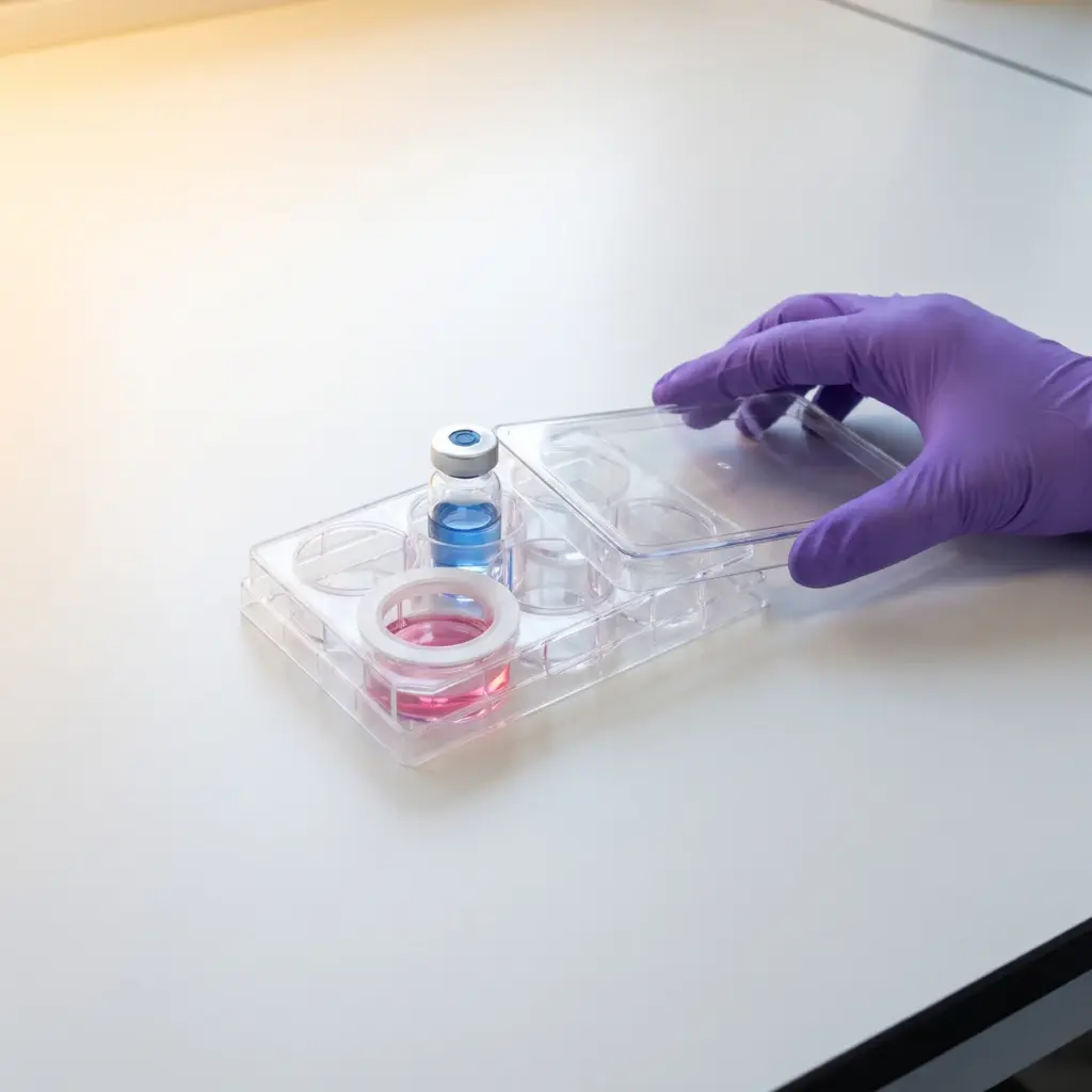
These studies compare treated and control skin-cell models after a specified challenge and read how much DNA damage forms and how it resolves. They characterise a defined mechanism in the cell model, which gives an ingredient claim a measured basis. Keratinocytes and fibroblasts are the usual cell types, chosen to match the skin layer the ingredient is meant to reach.
Measuring DNA damage
The core methods are single-cell gel electrophoresis, known as the comet assay, for strand breaks, and measurement of oxidative lesions such as 8-oxo-dG. The challenge can use selected particulate, ozone or metal exposures where they suit the model. Cells are treated before, during or after the challenge, depending on whether the question is reduced damage or faster recovery.
- DNA strand-break and related comet-assay endpoints
- Oxidative DNA-lesion measurements
- Comparison with challenge-only and unchallenged controls
- Cell viability to read damage signals in context
Investigating repair and recovery
Time-course designs show whether damage markers clear faster after treatment. Repair-protein expression and matched functional measurements help separate prevented damage from quicker recovery. Oxidative lesions such as 8-oxo-dG are cleared mainly by base excision repair, and bulkier lesions by nucleotide excision repair, so the study follows the pathway that matches the damage it induces. You receive the endpoint data, the control comparisons and a reading that names the mechanism actually tested, work that sits upstream of a pollutant protection claim.
Reading DNA protection mechanism findings
DNA data carries weight with ingredient buyers and formulators because it shows an effect at the level of the cell’s own genetic material. It is most useful when the study design and the claim are planned together from the start.
Choosing the right endpoint
Match the endpoint to the lesion or response you want to see. The comet assay and oxidative-lesion measurements report different things, and the exposure conditions shape the comparison. A lower initial damage signal can come from reduced exposure as much as from a cellular response, so the exposure has to be held constant across arms.
DNA damage recovery alongside stress response protein data
Recovery measurements show how damage changes over time, with viability and the starting damage level both weighing on any claim of protection or repair. Where the same active is proposed to lift the wider stress response, stress response protein testing adds a second line of mechanistic data. Set the decision criteria against your chosen model so results stay comparable across candidates.
More in Xenobiotic & Cellular Clearance Studies
Xenobiotic & Cellular Clearance Studies show how skin cells respond to pollutants and foreign compounds, and how an active supports their recycling system.