Clinical Imaging Microscopy overview
Do you provide Clinical Imaging Microscopy?
Clinical imaging microscopy methods for cosmetic studies
Each microscopy method looks at a different scale and depth, so the choice follows from the feature behind the claim.
Dermoscopy and video microscopy
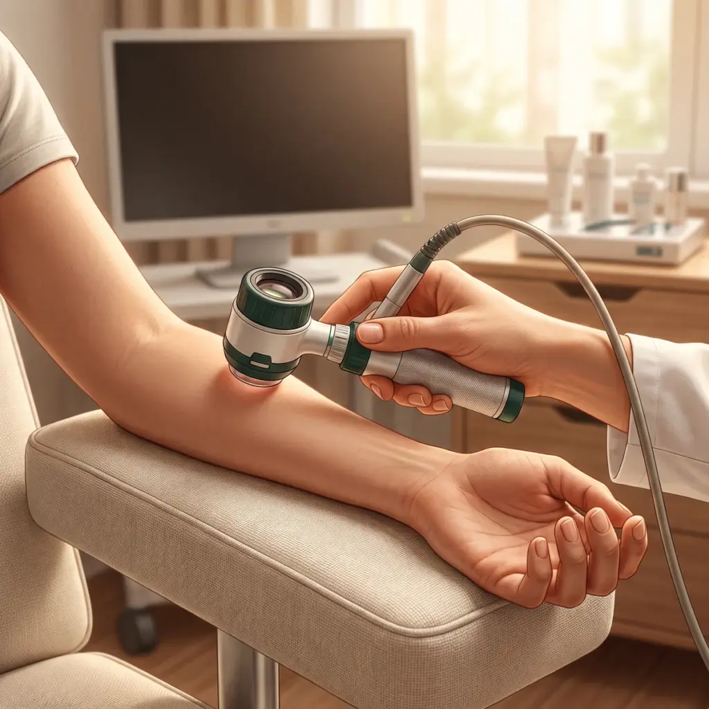
A dermoscope or handheld video microscope magnifies the skin surface under polarised or contact lighting. It records pore openings, follicles, scaling, fine surface lines and the pattern of superficial pigment. Images are quick to take and suit studies on pores, sebum plugs or surface smoothness.
Reflectance confocal microscopy
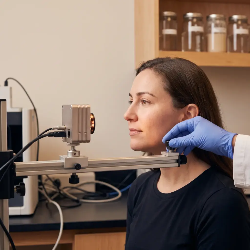
Reflectance confocal microscopy (RCM) scans a laser through the skin and collects reflected light from a single focal plane. The result is a stack of horizontal images at increasing depth through the epidermis to the upper dermis, at near-cellular resolution. Melanin reflects strongly, so RCM shows pigment distribution at the dermal-epidermal junction, along with keratinocyte layout and epidermal thickness. Optical coherence tomography (OCT) is a related method that gives vertical cross-sections over a wider area at lower resolution.
Histology from approved tissue sampling
Where a protocol includes ethically approved sampling, such as tape stripping or a small biopsy, prepared sections can be stained and examined by light microscopy for epidermal architecture, collagen-related features or selected markers.
Image capture and analysis
- Mark and record the study site at baseline
- Relocate the site with the same probe angle and pressure at each visit
- Capture image stacks at fixed depths and settings
- Grade or measure features within defined regions
- Compare time points statistically
Image stacks and pigment density tables delivered
You receive original image stacks, annotated example images, measurement tables (for example pigment density scores, pore counts or epidermal thickness) and a methods section ready for the study report.
Adding clinical imaging microscopy to an efficacy programme
Microscopy earns its place when you need to show what is happening beneath a visible result. A brightening product might pair cross-polarised photography of dark spots with RCM images showing a change in pigment at the dermal-epidermal junction. A pore-refining claim can use dermoscopy counts alongside photographs. The microscopy data supports mechanism wording and gives the visible claim a clear biological explanation.
Claim, body site and active data to share
Describe the claim and the feature you expect to change, the body site, the panel profile and the visit schedule. Say which method you have in mind or ask the laboratory to recommend one. Share any in vitro or ex vivo data on the active, since it helps the laboratory choose the depth and markers worth tracking.
Questions to ask an imaging microscopy provider
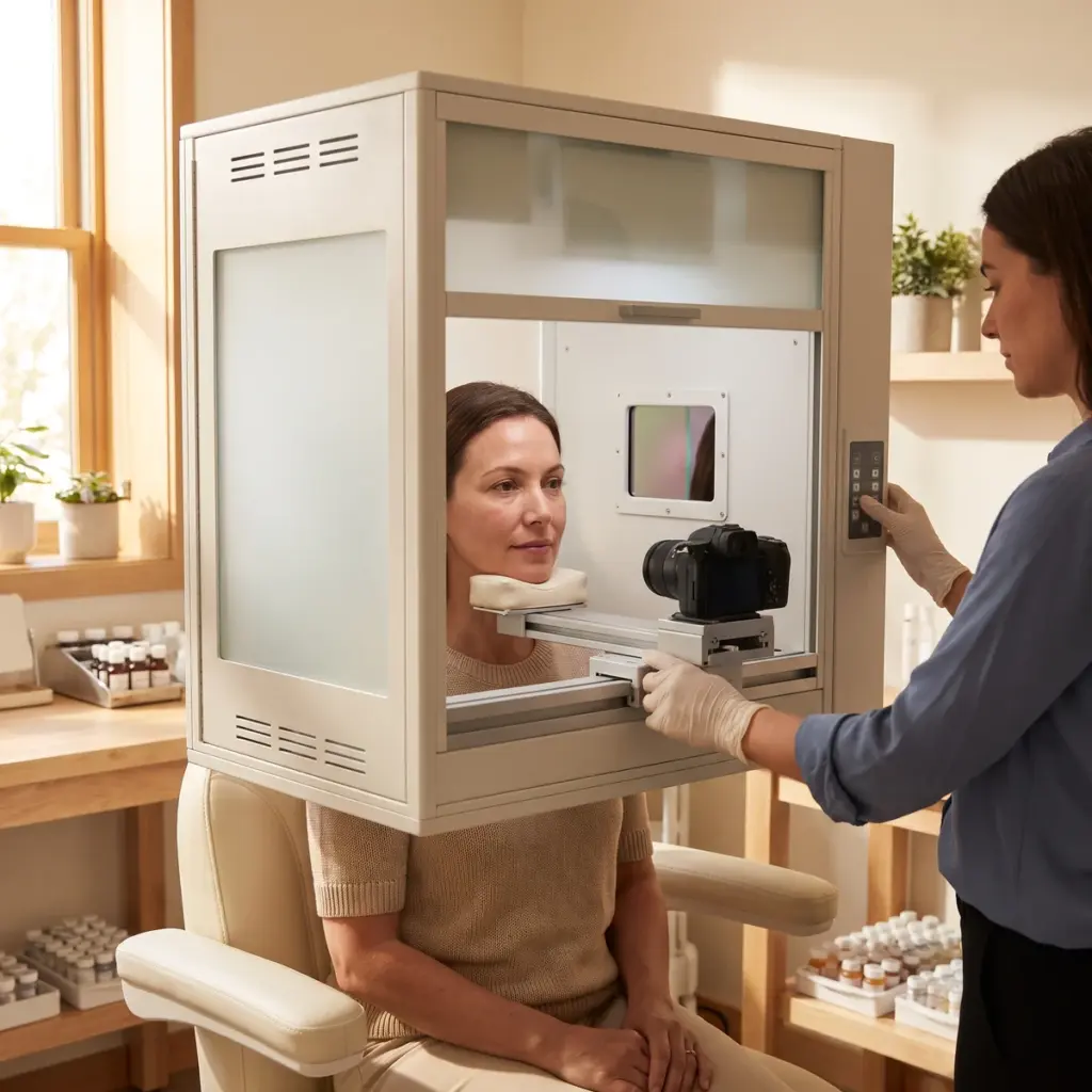
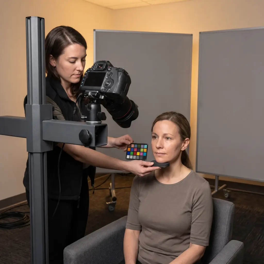
Ask which instruments they own, who operates them and how operators are trained to relocate the same site between visits. Request example image stacks and the scoring method used for pigment or structure. Check whether readers are blinded to visit and product. Confirm data formats and how images will be presented in the clinical study report. For whole-face evidence of the same change, pair the microscopy with standardised photography.
More in Clinical imaging documentation
Clinical imaging documentation records skin at each study visit with controlled photography, UV, 3D or microscopic imaging for claim evidence.