UV photography overview
Do you provide UV photography?
How UV photography reveals hidden skin features
Melanin absorbs ultraviolet light strongly. Under UV, areas with more epidermal pigment therefore appear darker against the surrounding skin, so freckles, sun spots and mottled patches that are faint in daylight show up as distinct dark areas. This contrast is the basis for following pigment changes during a brightening study.
Reflected UV and UV fluorescence
Reflected UV photography lights the skin with ultraviolet and fits the camera with a filter that passes only UV, recording the absorption pattern. Fluorescence photography lights the skin with UV and filters out the UV itself, recording only the visible light the skin emits. In fluorescence images, porphyrins produced by skin bacteria in pores glow orange-red, which allows pore occupancy to be counted before and after a cleansing or sebum-regulating product.
Visualising product on the skin
UV filters in sunscreens absorb ultraviolet, so a treated area looks dark in a reflected UV image. Laboratories use this to show the evenness of a sunscreen film, to compare application methods or to record how much product remains after water immersion or rubbing. Some cosmetic films and tracers fluoresce, which lets coverage be mapped the same way.
Capture controls
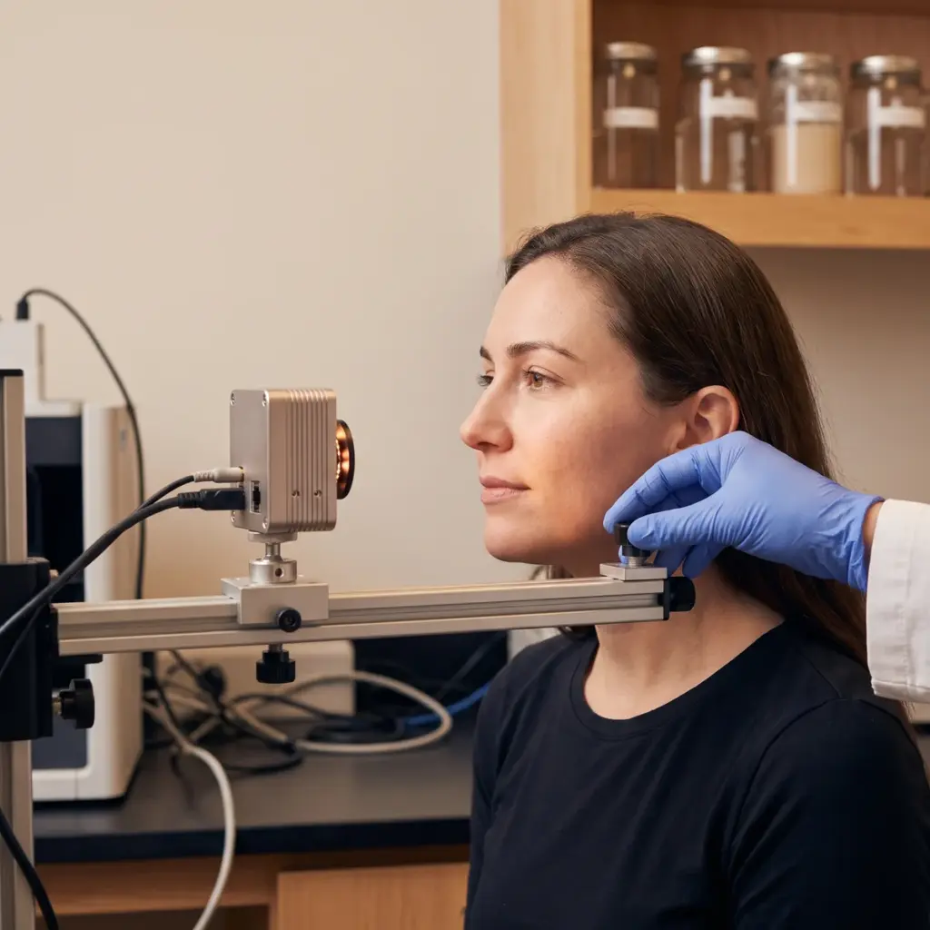
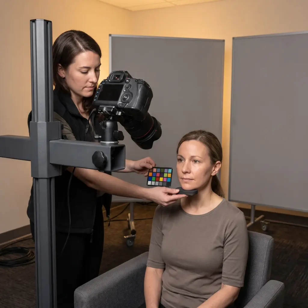
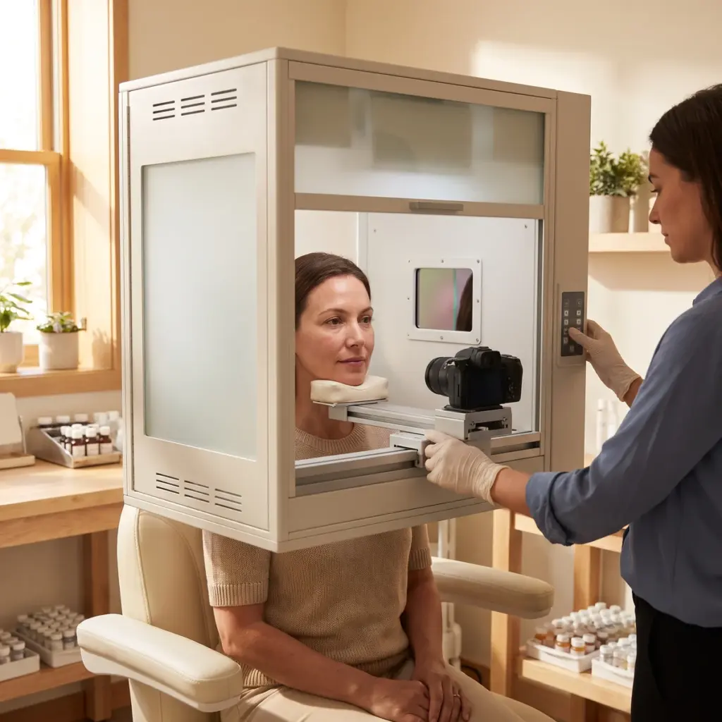
- Fixed head position and camera distance
- Same UV source, filters and exposure at every visit
- Skin cleansed and free of residue before capture
- Reference target in frame for exposure checks
- Defined analysis regions on each image
Outputs
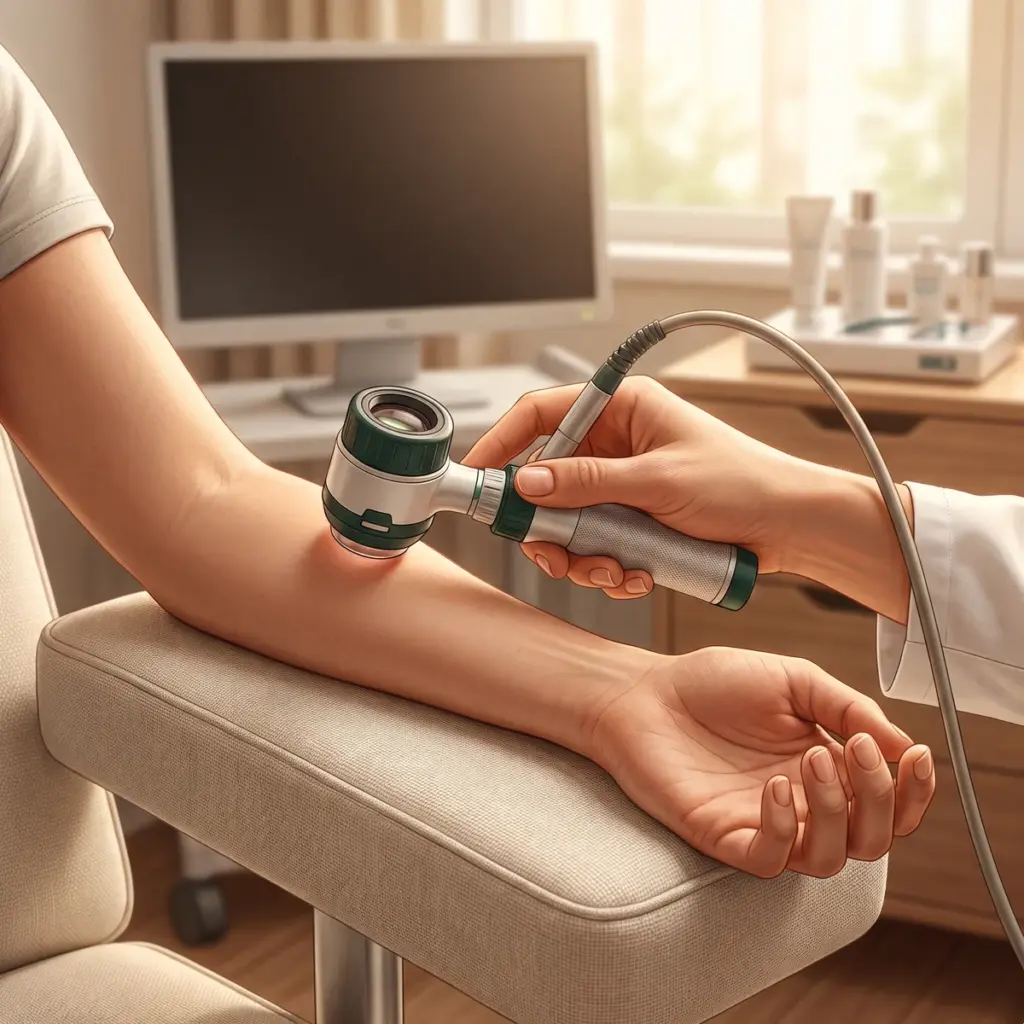
You receive original UV images with acquisition settings, matched visit sets, and analysis results such as spot count, spot area, pigment contrast or fluorescent pore count, ready for the study report.
Planning UV photography in a brightening or suncare study
UV photography fits claims about pigmentation, pores or product coverage. For an even-tone serum, reflected UV images taken at baseline and at each follow-up show pigment spots fading or contrast falling. For a pore-cleansing product, fluorescence images count porphyrin spots. For a sunscreen, reflected UV images show where the film is thin or missing, useful for application guidance and for claims about even coverage.
UV mode and marketing consent details to give
Name the claim, the area to image and whether you need reflected UV, fluorescence or both. Describe the product, its application amount and any water or rub resistance step for coverage studies. Say whether UV images will appear in marketing, since volunteers must consent to that use.
Selecting a UV imaging laboratory
Ask which UV sources and filters the system uses and how exposure is checked between sessions. Request example image sets from a previous study and the analysis method behind any spot or pore counts. Check that volunteers are positioned the same way each visit and that originals are kept with their metadata. For pigment evidence in normal light to accompany the UV series, add cross-polarised standardised photography at the same visits.
More in Clinical imaging documentation
Clinical imaging documentation records skin at each study visit with controlled photography, UV, 3D or microscopic imaging for claim evidence.