3D surface topography overview
Do you provide 3D surface topography?
How 3D surface topography measures skin relief
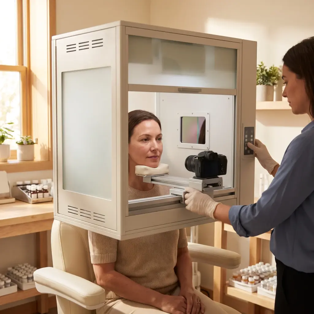
Most in vivo systems use fringe projection. A projector casts a series of striped light patterns onto the skin and a camera records how the stripes bend over the surface. Software converts the distortion into a height value for each point, building a 3D model of the area in a fraction of a second. Other systems reconstruct relief from images lit from several directions. Both suit the crow’s feet, forehead, nasolabial fold and cheek.
Replica profilometry
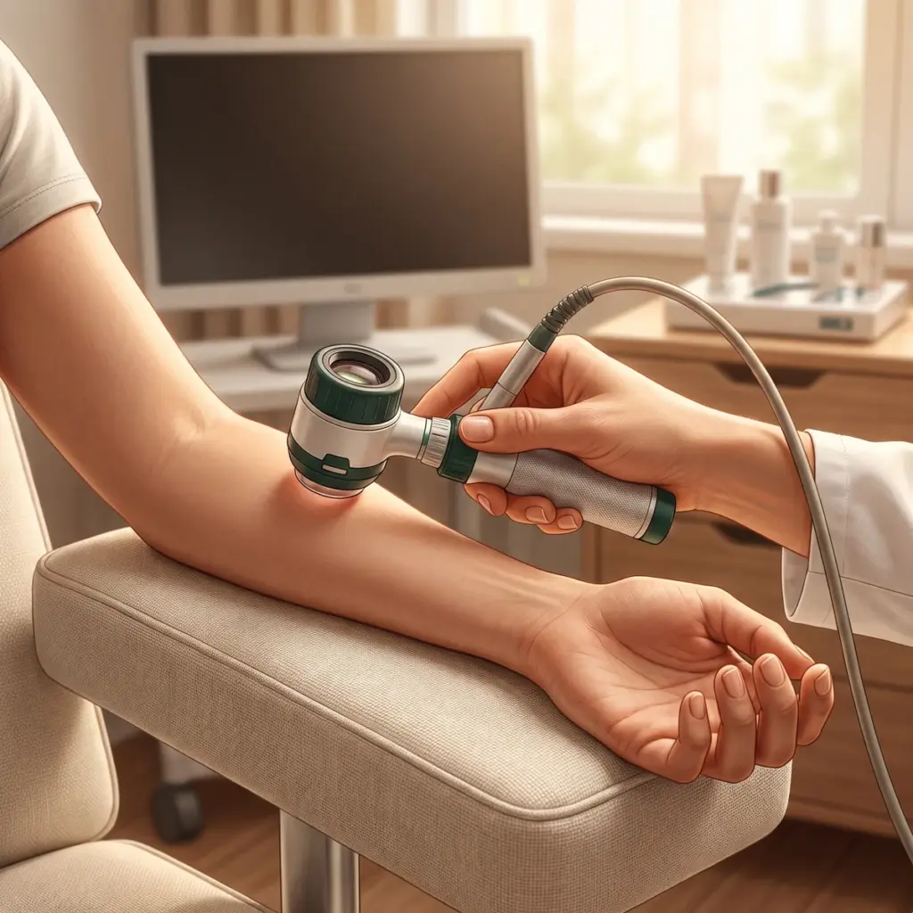
Some studies take a silicone impression of the skin at each visit and leave the face itself unscanned. The replica is then measured in the laboratory by optical or stylus profilometry. Replicas freeze the surface at the moment of sampling and can be stored and re-measured later.
Parameters reported
- Maximum and mean wrinkle depth
- Wrinkle volume and area
- Surface roughness values such as Ra and Rz
- Local volume or contour change in a defined zone
- Colour-coded height maps for each visit
Keeping scans comparable between visits
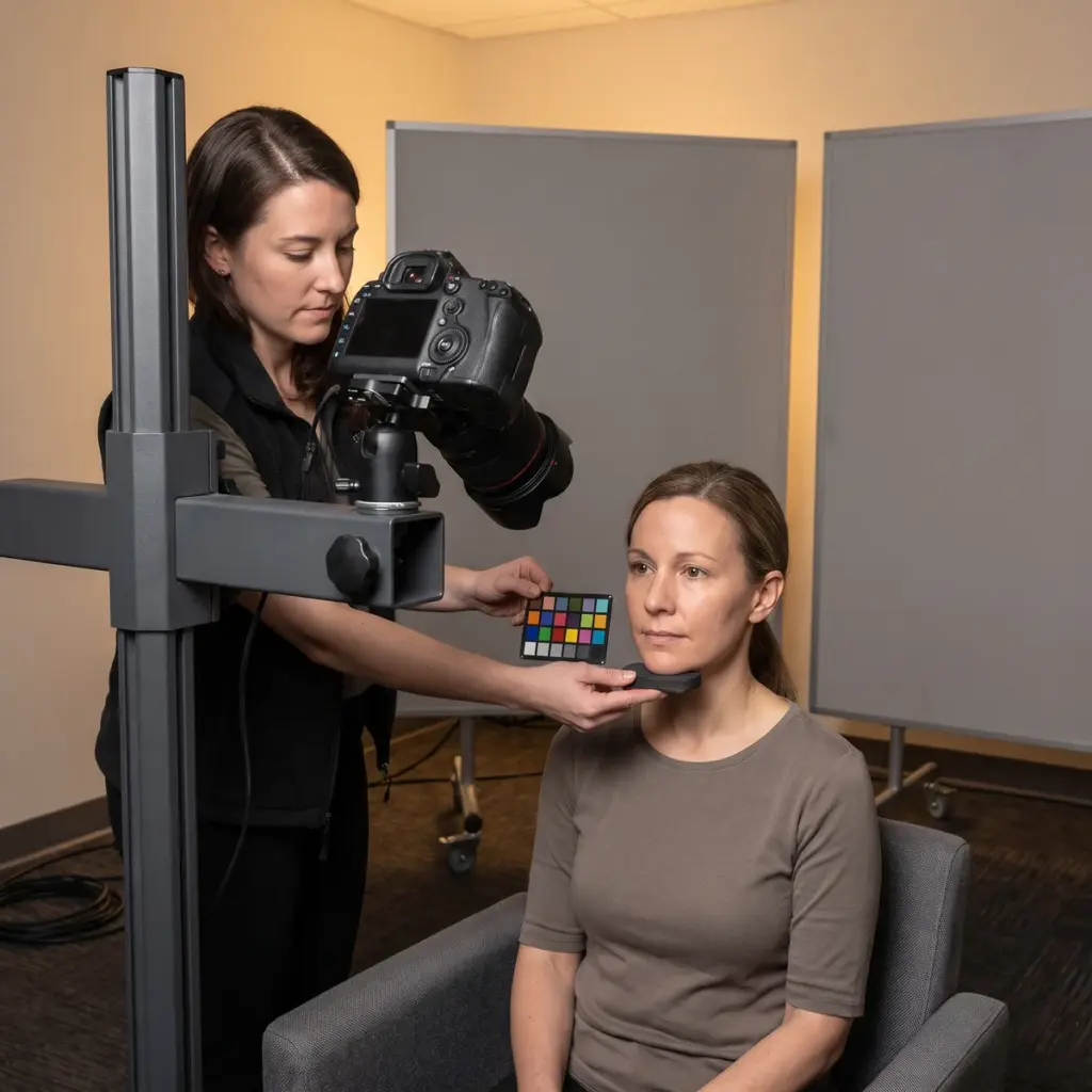
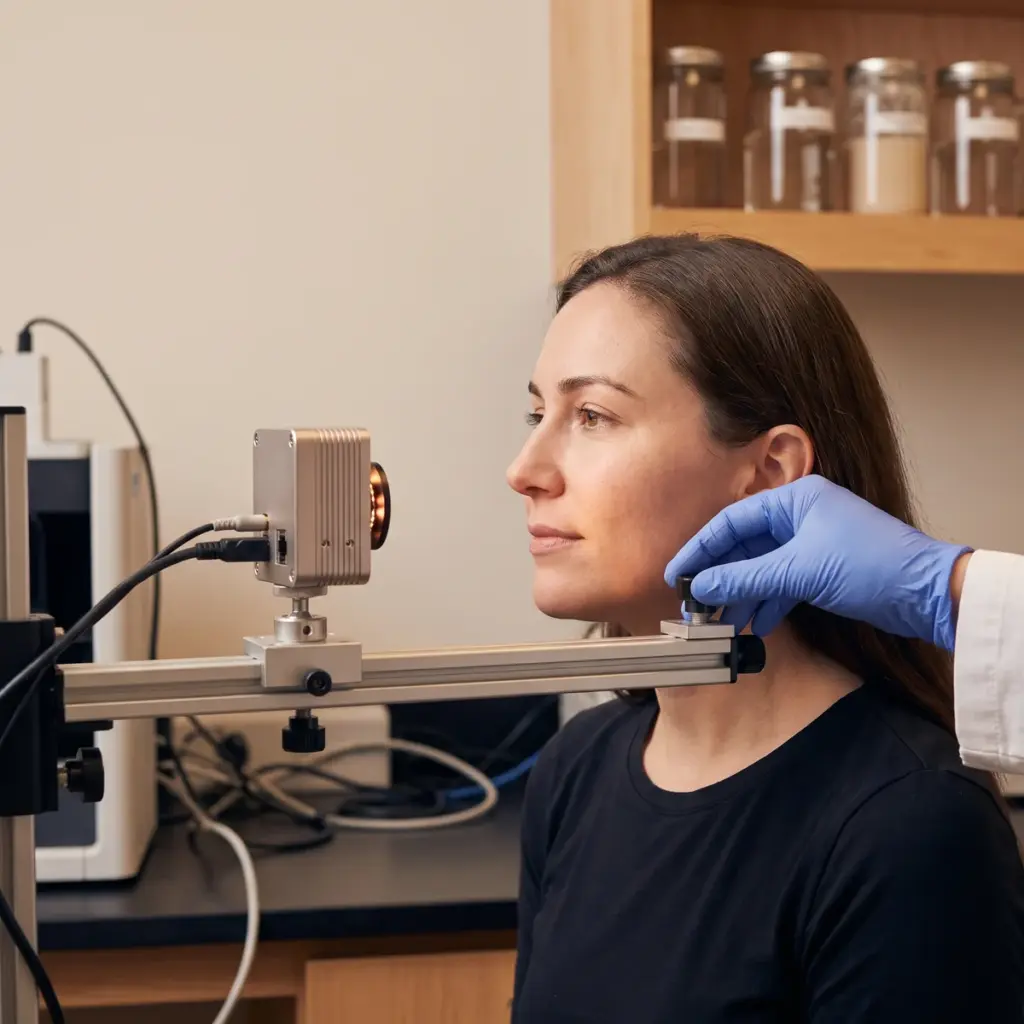
The volunteer is positioned with head supports and asked to hold a relaxed expression with eyes gently closed. The software overlays each follow-up scan on the baseline using matching algorithms, so the same patch of skin is analysed every time. Room temperature and humidity are controlled, and the system is checked against a calibration target.
Raw 3D files and height maps provided
The laboratory provides raw 3D files, the extracted parameters per volunteer and visit, statistical comparison against baseline or a control side, and paired height maps that show the change visually.
Using 3D surface topography to support anti-ageing claims
Choose topography when the claim is about the shape of the skin: fewer or shallower wrinkles, smoother texture, a fuller-looking lip or cheek, or reduced under-eye puffiness. It gives the statistical result that copy such as “wrinkle depth visibly reduced” needs, and the colour height maps make strong supporting visuals. For a perception claim on the same product, add a consumer questionnaire at the same visits.
Wrinkle zone and target parameter to specify
State the claim, the zone to measure and the product’s use routine. Give the visit schedule and whether a split-face or placebo design is planned. If you already have a target parameter in mind (depth, volume, roughness), say so, because it sets the analysis software settings and region size.
Picking a topography provider
Ask which scanner or replica method the laboratory uses and its stated resolution over the zone you care about. Request repeatability data from repeated scans of the same volunteer. Check how they register follow-up scans to baseline and how they handle volunteers whose expression changes. A provider that also runs standardised photography can capture both at each visit, so the wrinkle numbers and the pictures come from the same session.
More in Clinical imaging documentation
Clinical imaging documentation records skin at each study visit with controlled photography, UV, 3D or microscopic imaging for claim evidence.