Standardized photography overview
Do you provide Standardized photography?
Inside a standardized photography protocol
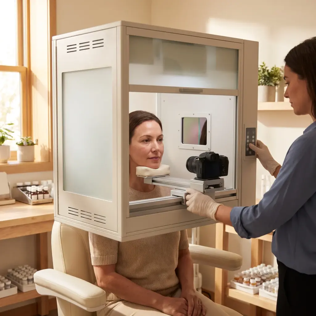
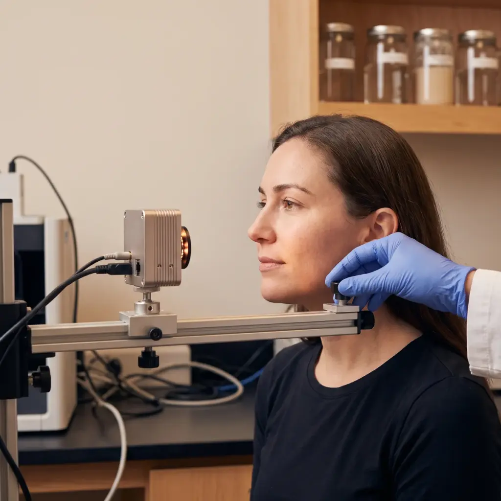
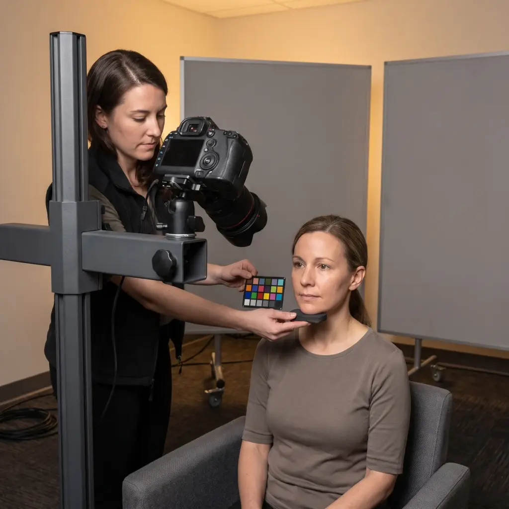
A capture booth fixes the volunteer’s head with a chin rest and forehead support, and a positioning overlay of the baseline image helps match the pose at later visits. Flash or continuous lights sit at fixed positions and intensities. Camera body, lens, aperture, shutter speed, white balance and distance stay the same for the whole study.
Lighting modes for different features
Standard diffuse lighting shows overall appearance. Cross-polarised light removes surface shine and brings out redness and brown pigmentation beneath the surface. Parallel-polarised light does the reverse, emphasising surface texture, fine lines and gloss. Many systems capture all three in one sitting, so graders have the right view for each endpoint.
Volunteer preparation and colour control
Volunteers arrive without make-up, cleanse at the centre and acclimatise to the room before capture. A neutral expression is coached, and hair is held back with a band. A colour reference chart in the frame or a calibration routine before each session keeps colour comparable across visits and cameras.
Grading and image analysis
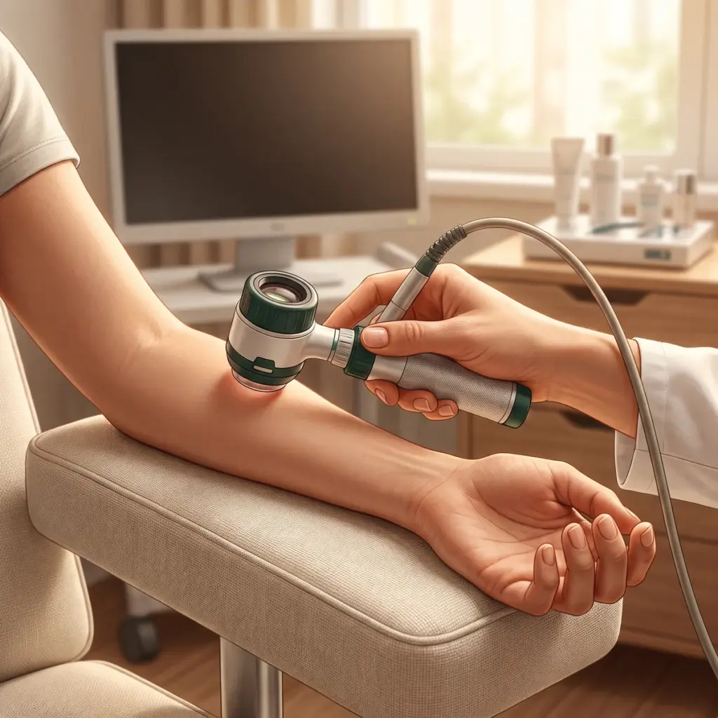
Expert graders score features against validated photonumeric scales, ideally blind to which visit each image comes from. Analysis software can measure spot count and area, redness index, pore size or skin tone uniformity within defined regions of interest. Both approaches produce numbers that go into the study statistics.
Files supplied at the end of the study
The laboratory delivers original unedited files with their metadata, matched image sets per volunteer and visit, grading scores or analysis outputs, and a capture procedure document. Images chosen for advertising are drawn from the same sets, so every marketing visual traces back to a study record.
Commissioning standardized photography for claim support
Plan photography into the protocol from the first draft. The claim determines the view (full face, profile, crow’s feet, décolletage, hands), the lighting mode and the grading scale. A visible-wrinkle claim might use parallel-polarised close-ups graded on a wrinkle scale. An even-tone claim might rely on cross-polarised full-face images analysed for pigment spots.
Briefing the imaging team
Send the claims, the body areas, the number of visits and whether images will appear in advertising. Marketing use needs specific consent from volunteers, so raise it before recruitment. State the grading scales or analysis outputs you expect and any retailer or market requirements on how before-and-after images may be shown.
What to check in a photography laboratory
Ask to see the capture booth and a set of baseline and follow-up images from a past study. Look at how closely pose and lighting match between visits. Confirm how often cameras and lights are calibrated, how graders are trained and whether they work blind. Check file retention terms and who owns the images. For deeper wrinkle measurements alongside photographs, add 3D surface topography to the same visits.
More in Clinical imaging documentation
Clinical imaging documentation records skin at each study visit with controlled photography, UV, 3D or microscopic imaging for claim evidence.