Analytical Microscopy overview
Do you provide Analytical Microscopy?
Microscopy techniques for cosmetic microstructure
Each microscope shows a different kind of feature, so the laboratory chooses the technique from the question you bring. Several can be combined on one sample.
Light and polarised light microscopy
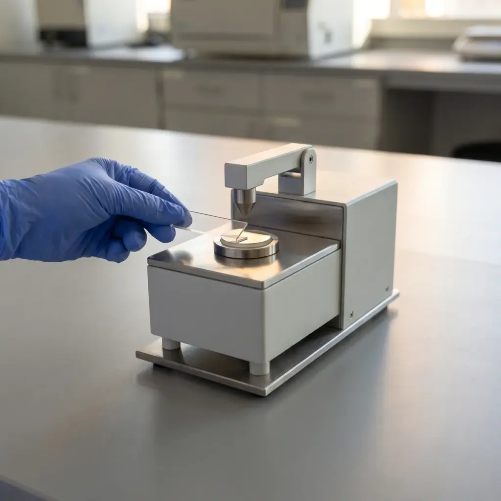
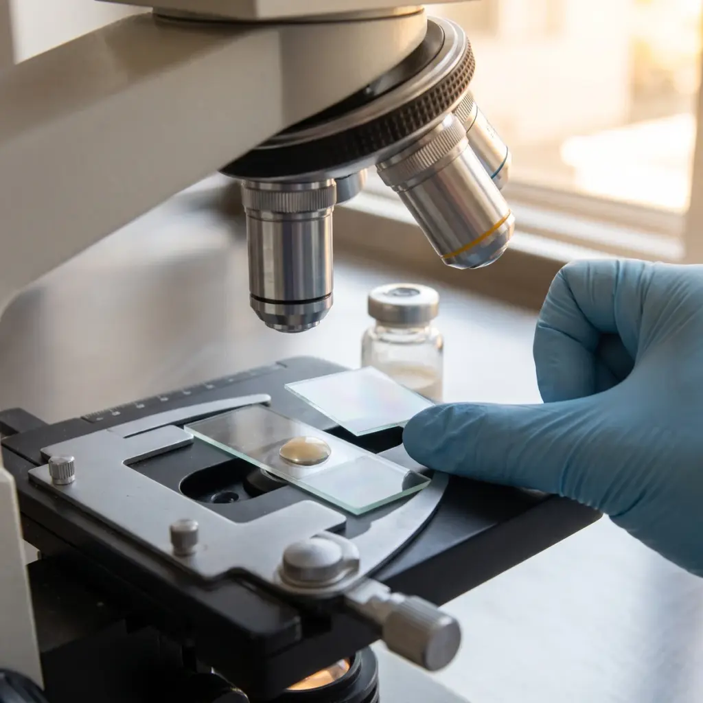
Bright-field light microscopy is the everyday tool for emulsions and dispersions. A thin film of product under a cover slip shows droplet size, uniformity and signs of flocculation or coalescence. Polarised light picks out birefringent features: crystals of fatty alcohols or actives growing out of a cream, and the lamellar liquid-crystalline phases that structure many emulsions.
Fluorescence and electron microscopy
Fluorescence microscopy locates components that fluoresce naturally or have been stained, for example to show which phase an ingredient sits in. Scanning electron microscopy gives high-magnification images of particle shape and surface, useful for pigments, mineral filters, exfoliants and encapsulates. Transmission electron microscopy resolves finer structures such as some vesicle systems. Electron microscopy usually needs drying or freezing, so the preparation is chosen to keep the feature of interest intact.
Sample preparation and image analysis
Dilution, drying, staining and the pressure of a cover slip can all change a soft sample. The laboratory records how each slide was made and keeps the method fixed for comparisons. Image analysis software can then measure droplet or particle size across many fields of view.
Micrographs, magnification and structure measurements delivered
The report contains representative micrographs with scale bars, the technique and magnification, a description of the structures seen and any quantitative measurements, with side-by-side panels when samples are compared.
Solving formulation problems with analytical microscopy
Most requests start with a sample that looks or feels wrong, or with two process options that need a clear winner.
Comparing emulsion prototypes
When the same formula is made at different homogenisation speeds, mixing times or temperatures, microscopy shows how the droplet population changes. In one published study of a cosmetic Pickering emulsion, slides of each sample revealed clear differences in droplet size and distribution across the mixing conditions. That kind of comparison lets you pick process settings with evidence before scale-up, and quantitative sizing from particle size distribution analysis can then put numbers on the trend.
Investigating graininess and streaking
A cream that turns gritty in storage often contains crystals, which polarised light shows at once. A foundation that streaks may carry pigment agglomerates that light microscopy picks up on a drawn-down film. Seeing the cause directly tells the formulator whether to change the wax blend, the cooling profile or the pigment dispersion step.
Choosing a microscopy provider
Ask which techniques the laboratory runs in-house and whether it has experience with soft, oily cosmetic samples, which behave differently from the hard materials many imaging labs handle. For particle shape and surface you need access to SEM. For emulsions, a light microscope with polarisers, a camera and image analysis software covers most needs.
More in Physicochemical Testing
Physicochemical testing measures a cosmetic’s active content, pH, viscosity, particle size and structure to set specifications and explain changes.