Lymphatic drainage overview
Do you provide Lymphatic drainage?
Lymphatic drainage testing from cell models to volunteers
Lymphatic capillaries have thin walls and overlapping endothelial cells that open to take up interstitial fluid. Larger collecting vessels then pump the lymph onward by rhythmic contraction. Drainage studies look at the lymphatic cells themselves and at the amount of fluid held in the skin and subcutaneous tissue.
Lymphatic endothelial cell assays
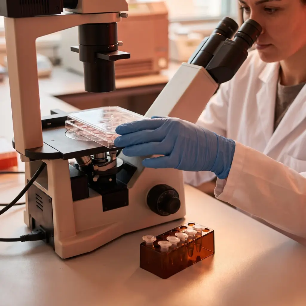
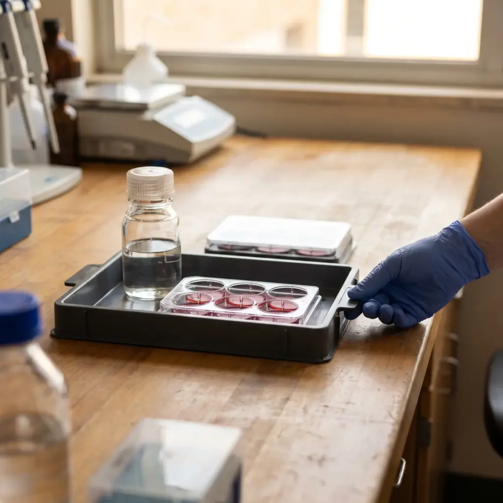
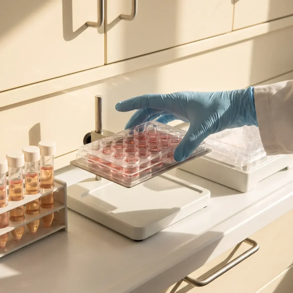
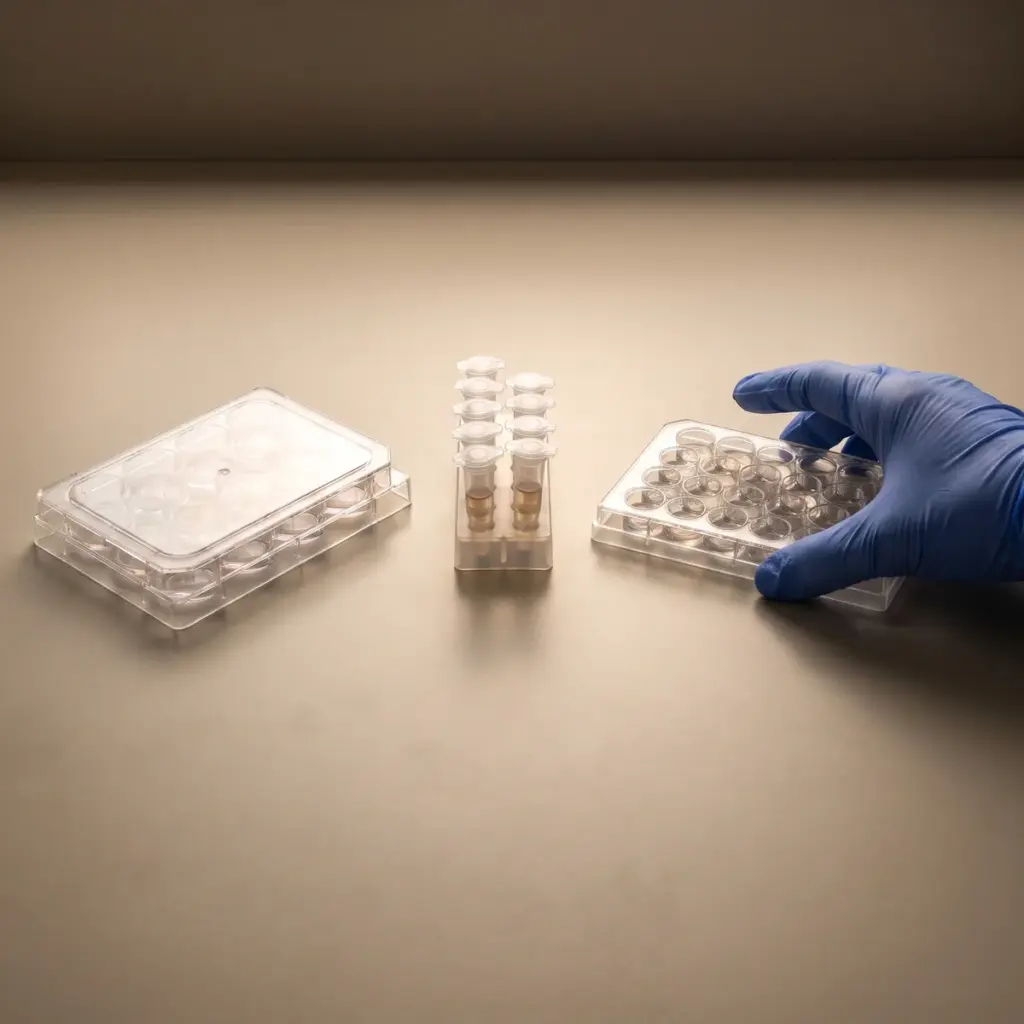
Human dermal lymphatic endothelial cells are cultured with the active and assessed for proliferation, migration and tube formation, which reflect the cells’ ability to build and maintain vessels. Expression of lymphatic markers such as PROX1, LYVE-1 and VEGFR-3 can be measured by qPCR. Monolayer permeability assays show whether the active changes the barrier between cells.
Fluid measurements on volunteers
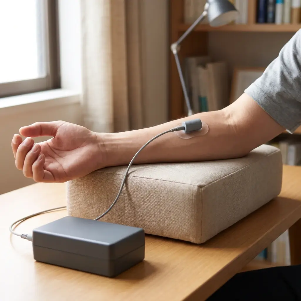
Clinical studies record tissue fluid at baseline and after a period of product use, usually on the legs or around the eyes. Typical endpoints are:
- local tissue water by bioimpedance or dielectric measurement
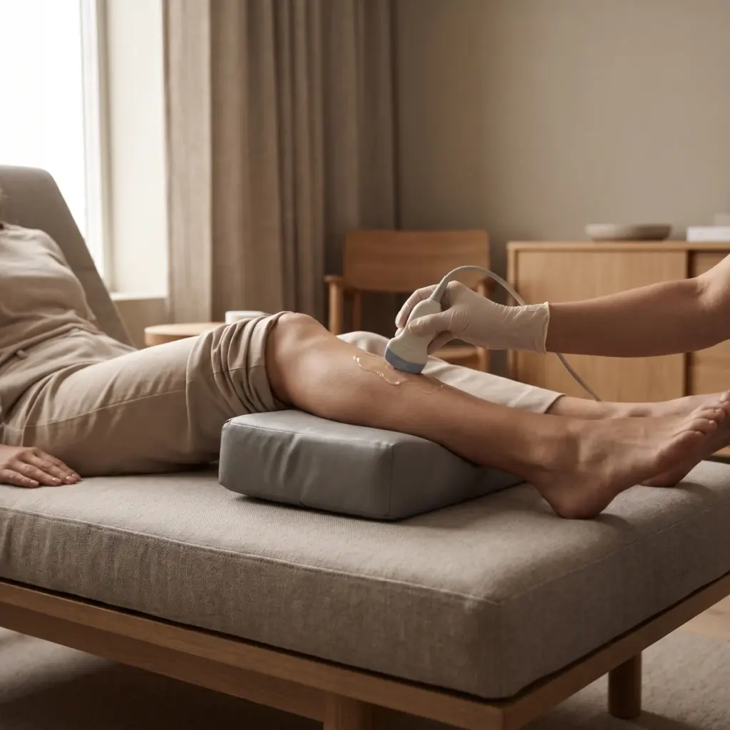
- dermal thickness and echogenicity by high-frequency ultrasound
- ankle and calf circumference or leg volume
- under-eye relief by 3D imaging
- volunteer ratings of heaviness and puffiness
Tube-network images and puffiness scores in the reports
Cell assay reports give each endpoint against vehicle and a reference growth factor, with images of tube networks. Clinical reports include the protocol, mean changes over time, statistics against baseline or a control zone and standardised images. Significant results support claims such as “visibly reduces under-eye puffiness” or “legs feel lighter”.
Matching lymphatic drainage testing to your product
The right endpoints depend on the body zone and on whether the claim is about how skin looks or how legs feel.
Eye-contour products
De-puffing eye gels and creams are usually tested on volunteers who wake with visible puffiness. Morning measurements by 3D imaging and ultrasound, repeated after application, capture the short-term effect. A use period of several weeks shows whether the improvement holds. Combining this with Microcirculation Assessment gives fuller dark-circle and puffiness evidence, since both blood and lymph flow affect the under-eye area.
Body and legs
For draining body gels and heavy-legs products, panels often include volunteers who stand for long periods, with measurements at the end of the day when swelling peaks. Leg circumference and bioimpedance give numbers for the claim, and volunteer questionnaires capture the feeling of lightness that consumers value. Anti-cellulite brands can fold these endpoints into a wider cellulite efficacy study.
Starting at the ingredient stage
Suppliers of draining actives typically begin with lymphatic endothelial assays to build a mechanism file. Tube formation and marker expression give formulators a clear picture of how the active works before a brand invests in a clinical study on the finished product.
More in Adipocyte & Vascular Studies
Adipocyte & Vascular Studies test body-care actives on fat-cell metabolism, lipolysis and skin microcirculation, in culture and on volunteers.