Sebocyte differentiation overview
Do you provide Sebocyte differentiation?
How sebocyte differentiation is measured in culture
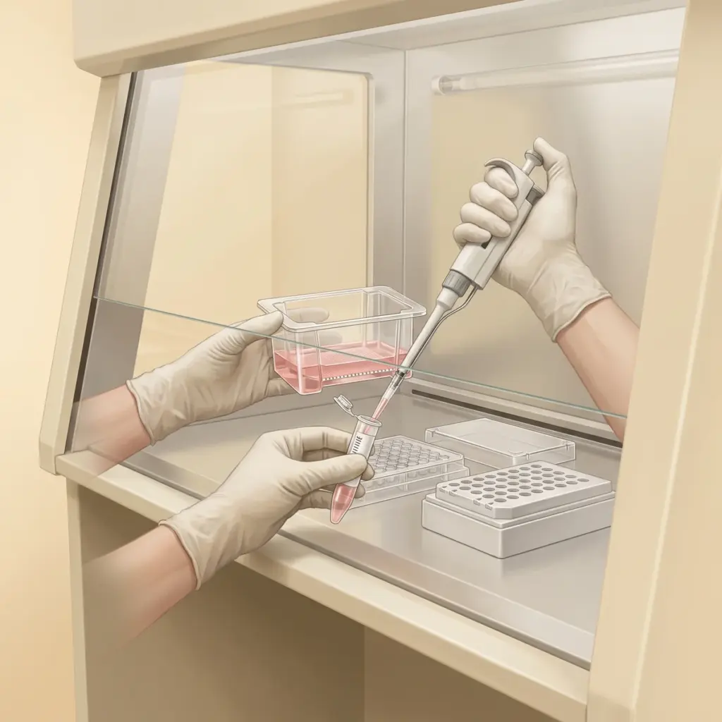
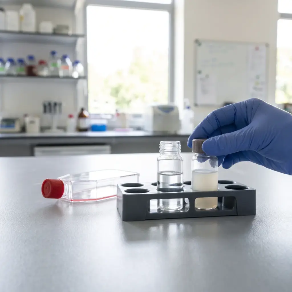
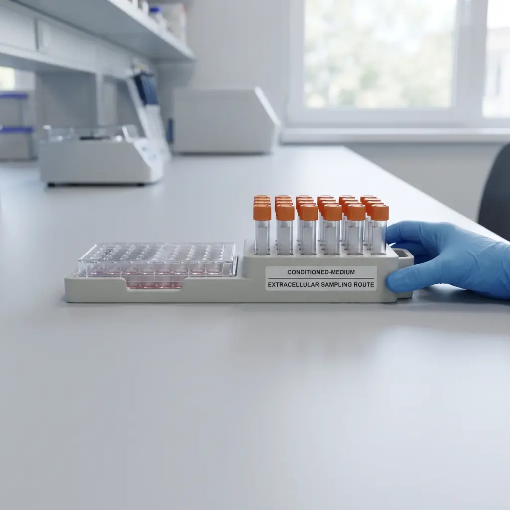
In the gland, sebocytes at the outer edge divide, move inward, accumulate lipid and finally break apart to release sebum. The assay reproduces the middle of that sequence in a dish. Cells are grown with a stimulus that pushes them to differentiate, such as linoleic acid or an androgen, and the test ingredient is added at several non-toxic concentrations alongside vehicle and reference wells.
Differentiation readouts
- cell size and morphology by phase-contrast microscopy
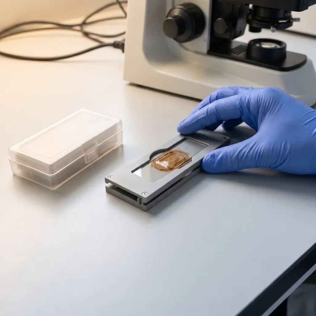
- lipid droplet number and area after Oil Red O or fluorescent staining
- PLIN2 and other maturation markers at gene or protein level
- lipogenic regulators such as SREBP-1 and PPARγ
- proliferation and viability in parallel wells
Choosing between SZ95 and primary sebocytes
The immortalised SZ95 line gives reproducible results across runs and is the usual choice for screening. Primary sebocytes from human donors are closer to the cells in skin and show donor-to-donor variation, which makes them useful for confirming a lead candidate. The laboratory times the readouts to the model, since the two mature at different speeds.
Separating maturation from cell number
Proliferation and viability counts run on the same plates. They show whether a lower lipid signal comes from fewer mature cells or from fewer cells in total. Results are normalised to cell number, so a change in the share of differentiated cells can be read directly.
Micrographs, droplet and marker data in the report
You receive representative micrographs for each condition, quantified droplet and marker data, concentration-response plots and the comparison with the reference compound.
When sebocyte differentiation data strengthens an oil-control claim
Sebocyte differentiation assays measure whether an active slows the maturation of sebocytes into lipid-filled cells. Order them when your positioning rests on that maturation step.
Positioning an active on cell maturation
Ingredient technical sheets often describe a mechanism such as balancing sebaceous activity or slowing sebocyte maturation. Differentiation data is the direct evidence for that wording: it shows fewer cells reaching the lipid-filled stage at a given concentration. It also gives formulators a reason to pair the active with ingredients that work further down the pathway.
Pairing with other sebocyte assays
Differentiation tells you whether cells become lipid producers. Lipogenesis inhibition tells you how much lipid those cells make. PPAR modulation often explains the link, because PPARγ signalling drives sebocyte differentiation. Ordering the three on the same model and dilution series produces one connected data set.
Differentiation stimulus, time points and SZ95 passage range
Ask which differentiation stimulus the laboratory uses and why, how many time points are included and whether markers are quantified at protein level by western blot or immunofluorescence. Check the passage range they use for SZ95 cells and which reference compound sets the benchmark.
More in Sebaceous Gland & Oily Skin
Sebaceous Gland & Oily Skin testing screens oil-control actives in sebocyte models for lipid production, androgen signalling and C. acnes activity.