Clinical 3D Imaging Documentation for Beauty Product Testing
What is Clinical 3D imaging documentation?
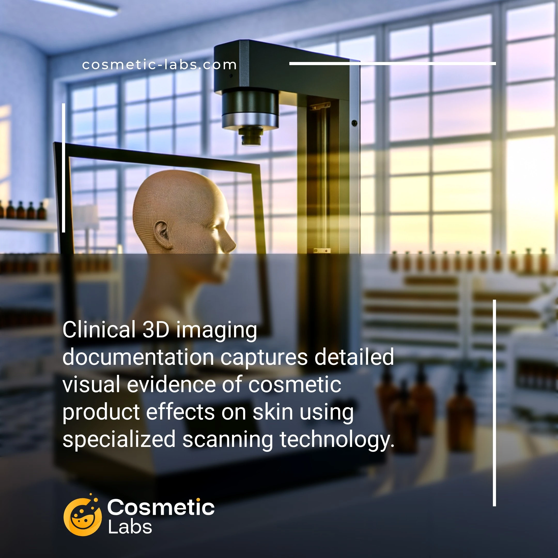
Clinical 3D imaging documentation services capture precise before-and-after visual evidence of cosmetic product efficacy through advanced volumetric scanning technology. Labs use specialized equipment like PRIMOS or Antera systems to measure skin texture changes, wrinkle depth reduction, and volume improvements with sub-millimeter accuracy. This documentation creates regulatory-compliant proof for anti-aging claims, supporting your product’s marketing statements with quantifiable data that meets FDA and international standards for cosmetic efficacy testing.
Why do you need this service?
Beauty brands leverage clinical 3D imaging documentation to capture before-and-after results for anti-aging serums, volumizing treatments, and skin texture improvements. Partner labs on our platform use advanced photogrammetry and volumetric analysis to document measurable changes in facial contours, wrinkle depth, and skin surface topology, delivering quantifiable proof of product efficacy that strengthens marketing claims and regulatory submissions.
Who provides Clinical 3D imaging documentation services?
All cosmetic labs providing Clinical 3D imaging documentation services
There is no company providing these services at the moment.
3D Imaging Documentation for Clinical Cosmetic Studies
3D imaging documentation captures detailed visual evidence of cosmetic product effects through precise volumetric measurements and surface analysis. Labs use this technology to create scientifically valid proof of your product’s performance claims, supporting regulatory submissions and marketing materials.
Advanced Imaging Protocols and Measurements
Professional cosmetic labs employ specialized 3D scanners that measure skin topography changes with micron-level precision. These systems track wrinkle depth, skin texture improvements, and volume changes over time.
Standard documentation includes:
- Baseline and follow-up 3D facial scans
- Quantitative before/after comparisons
- Statistical analysis of surface parameters
- Visual reports with color-coded mapping
Labs typically conduct imaging sessions at predetermined intervals—often at 2, 4, 8, and 12 weeks—to track progressive changes and establish efficacy timelines.
Clinical Study Integration and Regulatory Support
3D imaging data integrates seamlessly with clinical trials, providing objective measurements that support subjective assessments. This dual approach strengthens your product’s scientific credibility and regulatory compliance.
Documentation services include:
- Protocol development for imaging schedules
- Standardized lighting and positioning procedures
- Data analysis and statistical reporting
- Regulatory-compliant documentation packages
Connect with specialized labs on our platform to discuss 3D imaging requirements for your next clinical study and ensure your documentation meets industry standards.
Practical Applications of Clinical 3D Imaging Documentation
Clinical 3D imaging documentation services provide measurable proof of product efficacy through precise volumetric analysis and surface mapping technologies.
Anti-Aging Product Validation
Labs use 3D facial scanning systems like PRIMOS and Antera to capture wrinkle depth, skin texture, and volume changes before and after product application. These measurements track improvements in nasolabial folds, crow’s feet, and forehead lines with accuracy down to 0.1mm.
Documentation includes color-coded topographical maps showing surface elevation changes and statistical analysis of treated areas. Clinical studies typically run 4-12 weeks with imaging sessions every 2 weeks to establish efficacy timelines for marketing claims.
Body Contouring and Cellulite Treatment Documentation
3D body scanning captures circumference measurements, skin surface irregularities, and tissue density changes for slimming creams and anti-cellulite formulations. Labs measure thigh circumference, hip contours, and cellulite severity using standardized grading scales.
The documentation process involves controlled lighting conditions, standardized positioning protocols, and multiple angle captures. Results show percentage improvements in skin smoothness and measurable reductions in treated area circumferences, typically ranging from 2-8% over 8-week study periods.
| Measurement Type | Technology Used | Accuracy Level | Typical Study Duration |
|---|---|---|---|
| Wrinkle Depth Analysis | PRIMOS 3D Scanner | ±0.1mm | 4-12 weeks |
| Facial Volume Mapping | Antera 3D Camera | ±0.05mm | 6-16 weeks |
| Body Circumference | Fit3D Scanner | ±2mm | 8-12 weeks |
| Cellulite Severity | Optical Coherence Tomography | ±0.02mm | 6-10 weeks |
Ready to validate your cosmetic formulations with precise 3D imaging documentation? Contact specialized labs on our platform to discuss your clinical study requirements and establish measurable efficacy claims for your products.